 Movie
Movie Controller

[English] 日本語
 Yorodumi
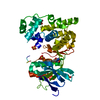
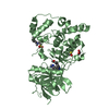
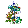
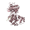
- PDB-4lub: X-ray structure of prephenate dehydratase from Streptococcus mutans -
+ Open data

- Basic information
| Entry | Database: PDB / ID: 4lub | ||||||
|---|---|---|---|---|---|---|---|
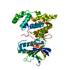
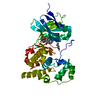
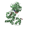
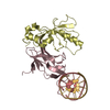
| Title | X-ray structure of prephenate dehydratase from Streptococcus mutans | ||||||
 Components Components | Putative prephenate dehydratase | ||||||
 Keywords Keywords | LYASE / dehydratase / dehydration | ||||||
| Function / homology |  Function and homology information Function and homology informationprephenate dehydratase / prephenate dehydratase activity / L-phenylalanine biosynthetic process / cytoplasm Similarity search - Function | ||||||
| Biological species |  Streptococcus mutans (bacteria) Streptococcus mutans (bacteria) | ||||||
| Method |  X-RAY DIFFRACTION / SYNCHROTRON / SAD / Resolution: 2.1 Å X-RAY DIFFRACTION / SYNCHROTRON / SAD / Resolution: 2.1 Å | ||||||
 Authors Authors | Shin, H.H. / Ku, H.K. / Song, J.S. / Choi, S. / Son, S.Y. | ||||||
 Citation Citation | Journal: TO BE PUBLISHED Title: X-ray structure of prephenate dehydratase from Streptococcus mutans Authors: Shin, H.H. / Ku, H.K. / Song, J.S. / Choi, S. / Son, S.Y. | ||||||
| History |
|
- Structure visualization
| Structure viewer | Molecule: MolmilJmol/JSmol |
|---|
- Downloads & links
-Download
| PDBx/mmCIF format | 4lub.cif.gz | 171.5 KB | Display | PDBx/mmCIF format |
|---|---|---|---|---|
| PDB format | pdb4lub.ent.gz | 135.9 KB | Display | PDB format |
| PDBx/mmJSON format | 4lub.json.gz | Tree view | PDBx/mmJSON format | |
| Others |  Other downloads Other downloads |
-Validation report
| Arichive directory | https://data.pdbj.org/pub/pdb/validation_reports/lu/4lubftp://data.pdbj.org/pub/pdb/validation_reports/lu/4lub | HTTPS FTP |
|---|
-Related structure data
| Similar structure data |
|---|


-Links
 PDBj
PDBj

- Assembly
| Deposited unit | 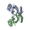
| ||||||||
|---|---|---|---|---|---|---|---|---|---|
| 1 |
| ||||||||
| Unit cell |
| ||||||||
| Components on special symmetry positions |
|
-Components
| #1: Protein | Mass: 31179.402 Da / Num. of mol.: 2 Source method: isolated from a genetically manipulated source Source: (gene. exp.) Streptococcus mutans (bacteria) / Strain: UA159 / Gene: pheA, SMU_786 / Plasmid: PET-28a / Production host: #2: Water | ChemComp-HOH / |  Mass: 18.015 Da / Num. of mol.: 228 / Source method: isolated from a natural source / Formula: H2O Mass: 18.015 Da / Num. of mol.: 228 / Source method: isolated from a natural source / Formula: H2O |
|---|
-Experimental details
-Experiment
| Experiment | Method: X-RAY DIFFRACTION / Number of used crystals: 1 |
|---|
- Sample preparation
| Crystal grow | Temperature: 293.15 K / Method: vapor diffusion, hanging drop / pH: 7.4 Details: 15% polyethylene glycol 8000, 100mM magnesium chloride, pH 7.4, VAPOR DIFFUSION, HANGING DROP, temperature 293.15K |
|---|
-Data collection
| Diffraction | Mean temperature: 100 K |
|---|---|
| Diffraction source | Source: SYNCHROTRON / Site: PAL/PLS  / Beamline: 6C1 / Wavelength: 1.2399 Å / Beamline: 6C1 / Wavelength: 1.2399 Å |
| Detector | Type: Photonic science VHR CCD / Detector: CCD / Date: Dec 6, 2009 |
| Radiation | Monochromator: DMM / Protocol: SINGLE WAVELENGTH / Monochromatic (M) / Laue (L): M / Scattering type: x-ray |
| Radiation wavelength | Wavelength: 1.2399 Å / Relative weight: 1 |
| Reflection | Resolution: 2.097→34.9 Å / Num. all: 25696 / Num. obs: 25692 / % possible obs: 99.6 % / Observed criterion σ(F): 1 / Observed criterion σ(I): 1 / Redundancy: 3.7 % / Biso Wilson estimate: 20.97 Å2 / Rmerge(I) obs: 0.0871 / Net I/σ(I): 14.86 |
| Reflection shell | Resolution: 2.1→2.18 Å / Redundancy: 3.7 % / Rmerge(I) obs: 0.0871 / Mean I/σ(I) obs: 14.86 / % possible all: 99.6 |
- Processing
| Software |
| ||||||||||||||||||||||||||||||||||||||||||||||||||||||||||||||||||||||
|---|---|---|---|---|---|---|---|---|---|---|---|---|---|---|---|---|---|---|---|---|---|---|---|---|---|---|---|---|---|---|---|---|---|---|---|---|---|---|---|---|---|---|---|---|---|---|---|---|---|---|---|---|---|---|---|---|---|---|---|---|---|---|---|---|---|---|---|---|---|---|---|
| Refinement | Method to determine structure: SAD / Resolution: 2.1→34.877 Å / SU ML: 0.21 / σ(F): 1.34 / Phase error: 23.07 / Stereochemistry target values: ML
| ||||||||||||||||||||||||||||||||||||||||||||||||||||||||||||||||||||||
| Solvent computation | Shrinkage radii: 0.9 Å / VDW probe radii: 1.11 Å / Solvent model: FLAT BULK SOLVENT MODEL | ||||||||||||||||||||||||||||||||||||||||||||||||||||||||||||||||||||||
| Refinement step | Cycle: LAST / Resolution: 2.1→34.877 Å
| ||||||||||||||||||||||||||||||||||||||||||||||||||||||||||||||||||||||
| Refine LS restraints |
| ||||||||||||||||||||||||||||||||||||||||||||||||||||||||||||||||||||||
| LS refinement shell |
| ||||||||||||||||||||||||||||||||||||||||||||||||||||||||||||||||||||||
| Refinement TLS params. | Method: refined / Origin x: 13.2841 Å / Origin y: 13.5826 Å / Origin z: 18.2263 Å
| ||||||||||||||||||||||||||||||||||||||||||||||||||||||||||||||||||||||
| Refinement TLS group | Selection details: ALL |
