| Entry | Database: PDB / ID: 4h3b
|
|---|
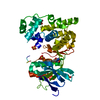
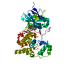
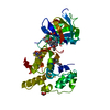
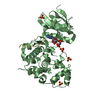
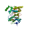
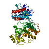
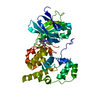
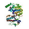
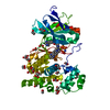
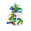
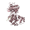
| Title | Crystal Structure of JNK3 in Complex with SAB Peptide |
|---|
 Components Components | - Mitogen-activated protein kinase 10
- SH3 domain-binding protein 5
|
|---|
 Keywords Keywords | TRANSFERASE / SH3BP-5 / MAPK / kinase |
|---|
| Function / homology |  Function and homology information Function and homology information
JUN kinase activity / Activation of the AP-1 family of transcription factors / Fc-epsilon receptor signaling pathway / MAP kinase kinase activity / protein kinase inhibitor activity / mitogen-activated protein kinase / response to light stimulus / JNK cascade / guanyl-nucleotide exchange factor activity / cytoplasmic vesicle membrane ...JUN kinase activity / Activation of the AP-1 family of transcription factors / Fc-epsilon receptor signaling pathway / MAP kinase kinase activity / protein kinase inhibitor activity / mitogen-activated protein kinase / response to light stimulus / JNK cascade / guanyl-nucleotide exchange factor activity / cytoplasmic vesicle membrane / JNK (c-Jun kinases) phosphorylation and activation mediated by activated human TAK1 / FCERI mediated MAPK activation / regulation of circadian rhythm / SH3 domain binding / cellular senescence / rhythmic process / Oxidative Stress Induced Senescence / protein phosphorylation / nuclear body / intracellular signal transduction / protein serine kinase activity / signal transduction / mitochondrion / nucleoplasm / ATP binding / nucleus / plasma membrane / cytosol / cytoplasmSimilarity search - Function SH3-binding 5 / SH3 domain-binding protein 5 (SH3BP5) / Mitogen-activated protein (MAP) kinase, JNK / Mitogen-activated protein (MAP) kinase, conserved site / MAP kinase signature. / : / Phosphorylase Kinase; domain 1 / Phosphorylase Kinase; domain 1 / Transferase(Phosphotransferase) domain 1 / Transferase(Phosphotransferase); domain 1 ...SH3-binding 5 / SH3 domain-binding protein 5 (SH3BP5) / Mitogen-activated protein (MAP) kinase, JNK / Mitogen-activated protein (MAP) kinase, conserved site / MAP kinase signature. / : / Phosphorylase Kinase; domain 1 / Phosphorylase Kinase; domain 1 / Transferase(Phosphotransferase) domain 1 / Transferase(Phosphotransferase); domain 1 / Serine/threonine-protein kinase, active site / Serine/Threonine protein kinases active-site signature. / Protein kinase domain / Serine/Threonine protein kinases, catalytic domain / Protein kinase domain profile. / Protein kinase domain / Protein kinase-like domain superfamily / 2-Layer Sandwich / Orthogonal Bundle / Mainly Alpha / Alpha BetaSimilarity search - Domain/homology |
|---|
| Biological species |  Homo sapiens (human) Homo sapiens (human) |
|---|
| Method |  X-RAY DIFFRACTION / SYNCHROTRON / MOLECULAR REPLACEMENT / Resolution: 2.08 Å X-RAY DIFFRACTION / SYNCHROTRON / MOLECULAR REPLACEMENT / Resolution: 2.08 Å |
|---|
 Authors Authors | Nwachukwu, J.C. / Laughlin, J.D. / Figuera-Losada, M. / Cherry, L. / Nettles, K.W. / LoGrasso, P.V. |
|---|
 Citation Citation | Journal: Structure / Year: 2012
Title: Structural Mechanisms of Allostery and Autoinhibition in JNK Family Kinases.
Authors: Laughlin, J.D. / Nwachukwu, J.C. / Figuera-Losada, M. / Cherry, L. / Nettles, K.W. / Lograsso, P.V. |
|---|
| History | | Deposition | Sep 13, 2012 | Deposition site: RCSB / Processing site: RCSB |
|---|
| Revision 1.0 | Nov 21, 2012 | Provider: repository / Type: Initial release |
|---|
| Revision 1.1 | Nov 28, 2012 | Group: Database references |
|---|
| Revision 1.2 | Dec 26, 2012 | Group: Database references |
|---|
| Revision 1.3 | Jun 17, 2015 | Group: Structure summary |
|---|
| Revision 1.4 | Sep 20, 2023 | Group: Data collection / Database references / Refinement description
Category: chem_comp_atom / chem_comp_bond ...chem_comp_atom / chem_comp_bond / database_2 / pdbx_initial_refinement_model
Item: _database_2.pdbx_DOI / _database_2.pdbx_database_accession |
|---|
|
|---|
 Movie
Movie Controller


 Open data
 Basic information
 Structure visualization
 Downloads & links
 Other downloads


 PDBj
PDBj


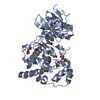
 Assembly
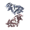


 Mass: 18.015 Da / Num. of mol.: 445 / Source method: isolated from a natural source / Formula: H2O
 Sample preparation
 / Beamline: BL11-1 / Wavelength: 1.17 Å
 Processing