 Movie
Movie Controller

[English] 日本語
 Yorodumi
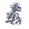
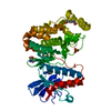
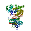
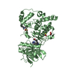
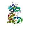
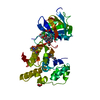
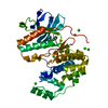
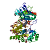
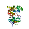
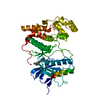
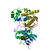
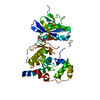
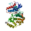
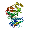
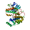
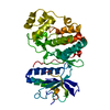
- PDB-1pmn: Crystal structure of JNK3 in complex with an imidazole-pyrimidine... -
+ Open data

- Basic information
| Entry | Database: PDB / ID: 1pmn | ||||||
|---|---|---|---|---|---|---|---|
| Title | Crystal structure of JNK3 in complex with an imidazole-pyrimidine inhibitor | ||||||
 Components Components | Mitogen-activated protein kinase 10 | ||||||
 Keywords Keywords | TRANSFERASE / MAP kinase / apoptosis / inhibition | ||||||
| Function / homology |  Function and homology information Function and homology informationJUN kinase activity / Activation of the AP-1 family of transcription factors / Fc-epsilon receptor signaling pathway / MAP kinase kinase activity / mitogen-activated protein kinase / response to light stimulus / JNK cascade / JNK (c-Jun kinases) phosphorylation and activation mediated by activated human TAK1 / FCERI mediated MAPK activation / regulation of circadian rhythm ...JUN kinase activity / Activation of the AP-1 family of transcription factors / Fc-epsilon receptor signaling pathway / MAP kinase kinase activity / mitogen-activated protein kinase / response to light stimulus / JNK cascade / JNK (c-Jun kinases) phosphorylation and activation mediated by activated human TAK1 / FCERI mediated MAPK activation / regulation of circadian rhythm / cellular senescence / rhythmic process / Oxidative Stress Induced Senescence / protein phosphorylation / protein serine kinase activity / signal transduction / mitochondrion / nucleoplasm / ATP binding / nucleus / plasma membrane / cytoplasm / cytosol Similarity search - Function | ||||||
| Biological species |  Homo sapiens (human) Homo sapiens (human) | ||||||
| Method |  X-RAY DIFFRACTION / SYNCHROTRON / FOURIER SYNTHESIS / Resolution: 2.2 Å X-RAY DIFFRACTION / SYNCHROTRON / FOURIER SYNTHESIS / Resolution: 2.2 Å | ||||||
 Authors Authors | Scapin, G. / Patel, S.B. / Lisnock, J. / Becker, J.W. / LoGrasso, P.V. | ||||||
 Citation Citation | Journal: Chem.Biol. / Year: 2003 Title: The structure of JNK3 in complex with small molecule inhibitors: structural basis for potency and selectivity Authors: Scapin, G. / Patel, S.B. / Lisnock, J. / Becker, J.W. / LoGrasso, P.V. | ||||||
| History |
|
- Structure visualization
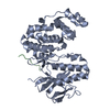
| Structure viewer | Molecule: MolmilJmol/JSmol |
|---|
- Downloads & links
-Download
| PDBx/mmCIF format | 1pmn.cif.gz | 88.7 KB | Display | PDBx/mmCIF format |
|---|---|---|---|---|
| PDB format | pdb1pmn.ent.gz | 64.9 KB | Display | PDB format |
| PDBx/mmJSON format | 1pmn.json.gz | Tree view | PDBx/mmJSON format | |
| Others |  Other downloads Other downloads |
-Validation report
| Arichive directory | https://data.pdbj.org/pub/pdb/validation_reports/pm/1pmnftp://data.pdbj.org/pub/pdb/validation_reports/pm/1pmn | HTTPS FTP |
|---|
-Related structure data
| Related structure data |  1pmuC  1pmvC  4z9lC  1jnkS C: citing same article ( S: Starting model for refinement |
|---|---|
| Similar structure data |


-Links
 PDBj
PDBj


- Assembly
| Deposited unit | 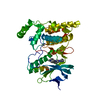
| ||||||||
|---|---|---|---|---|---|---|---|---|---|
| 1 |
| ||||||||
| Unit cell |
|
-Components
| #1: Protein | Mass: 42001.641 Da / Num. of mol.: 1 Source method: isolated from a genetically manipulated source Source: (gene. exp.) Homo sapiens (human) / Gene: MK10_HUMAN / Plasmid: pET15B / Production host:  References: UniProt: P53779, Transferases; Transferring phosphorus-containing groups; Phosphotransferases with an alcohol group as acceptor |
|---|---|
| #2: Chemical | ChemComp-984 / 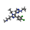  Mass: 485.452 Da / Num. of mol.: 1 / Source method: obtained synthetically / Formula: C25H30Cl2N6 Mass: 485.452 Da / Num. of mol.: 1 / Source method: obtained synthetically / Formula: C25H30Cl2N6 |
| #3: Water | ChemComp-HOH /  Mass: 18.015 Da / Num. of mol.: 197 / Source method: isolated from a natural source / Formula: H2O Mass: 18.015 Da / Num. of mol.: 197 / Source method: isolated from a natural source / Formula: H2O |
-Experimental details
-Experiment
| Experiment | Method: X-RAY DIFFRACTION / Number of used crystals: 1 |
|---|
- Sample preparation
| Crystal | Density Matthews: 2.41 Å3/Da / Density % sol: 49.05 % | |||||||||||||||||||||||||||||||||||||||||||||||||
|---|---|---|---|---|---|---|---|---|---|---|---|---|---|---|---|---|---|---|---|---|---|---|---|---|---|---|---|---|---|---|---|---|---|---|---|---|---|---|---|---|---|---|---|---|---|---|---|---|---|---|
| Crystal grow | Temperature: 293 K / Method: vapor diffusion, hanging drop / pH: 7 Details: PEG MMe 550, Ethylene glycol, Hepes, pH 7.0, VAPOR DIFFUSION, HANGING DROP, temperature 293K | |||||||||||||||||||||||||||||||||||||||||||||||||
| Crystal grow | *PLUS pH: 7.3 / Method: vapor diffusion, hanging drop | |||||||||||||||||||||||||||||||||||||||||||||||||
| Components of the solutions | *PLUS
|
-Data collection
| Diffraction | Mean temperature: 100 K |
|---|---|
| Diffraction source | Source: SYNCHROTRON / Site: APS  / Beamline: 17-ID / Wavelength: 1 Å / Beamline: 17-ID / Wavelength: 1 Å |
| Detector | Type: MARRESEARCH / Detector: CCD / Date: Mar 6, 2000 |
| Radiation | Protocol: SINGLE WAVELENGTH / Monochromatic (M) / Laue (L): M / Scattering type: x-ray |
| Radiation wavelength | Wavelength: 1 Å / Relative weight: 1 |
| Reflection | Resolution: 2.2→22 Å / Num. all: 21324 / Num. obs: 21026 / % possible obs: 98.6 % / Observed criterion σ(F): 0 / Observed criterion σ(I): 0 / Redundancy: 6.1 % / Biso Wilson estimate: 30.9 Å2 / Rsym value: 0.059 / Net I/σ(I): 8.6 |
| Reflection shell | Resolution: 2.2→2.3 Å / Redundancy: 5.8 % / Mean I/σ(I) obs: 2.4 / Num. unique all: 3483 / Rsym value: 0.197 / % possible all: 98.5 |
| Reflection | *PLUS Rmerge(I) obs: 0.059 |
| Reflection shell | *PLUS % possible obs: 98.5 % / Num. unique obs: 3431 / Rmerge(I) obs: 0.197 |
- Processing
| Software |
| ||||||||||||||||||||||||||||||||||||
|---|---|---|---|---|---|---|---|---|---|---|---|---|---|---|---|---|---|---|---|---|---|---|---|---|---|---|---|---|---|---|---|---|---|---|---|---|---|
| Refinement | Method to determine structure: FOURIER SYNTHESIS Starting model: PDB ENTRY 1JNK Resolution: 2.2→22 Å / Isotropic thermal model: Restrained / Cross valid method: THROUGHOUT / σ(F): 0 / σ(I): 0 / Stereochemistry target values: Engh & Huber
| ||||||||||||||||||||||||||||||||||||
| Solvent computation | Solvent model: Mask / Bsol: 52.9 Å2 / ksol: 0.396 e/Å3 | ||||||||||||||||||||||||||||||||||||
| Displacement parameters | Biso mean: 34.9 Å2
| ||||||||||||||||||||||||||||||||||||
| Refine analyze |
| ||||||||||||||||||||||||||||||||||||
| Refinement step | Cycle: LAST / Resolution: 2.2→22 Å
| ||||||||||||||||||||||||||||||||||||
| Refine LS restraints |
| ||||||||||||||||||||||||||||||||||||
| LS refinement shell | Resolution: 2.2→2.28 Å
| ||||||||||||||||||||||||||||||||||||
| Refine LS restraints | *PLUS
|
