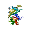
 Movie
Movie Controller


+ Open data

- Basic information
| Entry | Database: PDB / ID: 5rsa | |||||||||
|---|---|---|---|---|---|---|---|---|---|---|
| Title | COMPARISON OF TWO INDEPENDENTLY REFINED MODELS OF RIBONUCLEASE-A | |||||||||
 Components Components | RIBONUCLEASE A | |||||||||
 Keywords Keywords | Hydrolase/RNA / HYDROLASE-RNA Complex | |||||||||
| Function / homology |  Function and homology information Function and homology informationpancreatic ribonuclease / ribonuclease A activity / RNA nuclease activity / nucleic acid binding / defense response to Gram-positive bacterium / hydrolase activity / extracellular region Similarity search - Function | |||||||||
| Biological species |  | |||||||||
| Method |  X-RAY DIFFRACTION / NEUTRON DIFFRACTION / Resolution: 2 Å X-RAY DIFFRACTION / NEUTRON DIFFRACTION / Resolution: 2 Å | |||||||||
 Authors Authors | Wlodawer, A. | |||||||||
 Citation Citation | Journal: Acta Crystallogr.,Sect.B / Year: 1986 Title: Comparison of Two Independently Refined Models of Ribonuclease-A Authors: Wlodawer, A. / Borkakoti, N. / Moss, D.S. / Howlin, B. #1: Journal: Biochemistry / Year: 1985Title: Nuclear Magnetic Resonance and Neutron Diffraction Studies of the Complex of Ribonucleasea with Uridine Vanadate, a Transition-State Analogue Authors: Borah, B. / Chen, C.-W. / Egan, W. / Miller, M. / Wlodawer, A. / Cohen, J.S. #2: Journal: Biochemistry / Year: 1983Title: Structure of Ribonuclease A. Results of Joint Neutron and X-Ray Refinement at 2.0-Angstroms Resolution Authors: Wlodawer, A. / Sjolin, L. #3: Journal: Proc.Natl.Acad.Sci.USA / Year: 1983Title: Active Site of Rnase. Neutron Diffraction Study of a Complex with Uridine Vanadate, a Transition-State Analog Authors: Wlodawer, A. / Miller, M. / Sjolin, L. #4: Journal: J.Biol.Chem. / Year: 1982Title: The Refined Crystal Structure of Ribonuclease A at 2.0 Angstroms Resolution Authors: Wlodawer, A. / Bott, R. / Sjolin, L. #5: Journal: Proc.Natl.Acad.Sci.USA / Year: 1982Title: Hydrogen Exchange in Rnase A. Neutron Diffraction Study Authors: Wlodawer, A. / Sjolin, L. #6: Journal: Acta Crystallogr.,Sect.A (Supplement) / Year: 1981Title: Structure of Ribonuclease A. X-Ray and Neutron Refinement Authors: Wlodawer, A. / Bott, R. / Sjolin, L. #7: Journal: Acta Crystallogr.,Sect.A (Supplement) / Year: 1981Title: Joint Refinement of Macromolecular Structures with X-Ray and Neutron Single-Crystal Diffraction Data Authors: Wlodawer, A. / Hendrickson, W.A. #8: Journal: Proc.Natl.Acad.Sci.USA / Year: 1981Title: Orientation of Histidine Residues in Rnase A. Neutron Diffraction Study Authors: Wlodawer, A. / Sjolin, L. #9: Journal: Acta Crystallogr.,Sect.B / Year: 1980Title: Studies of Ribonuclease-A by X-Ray and Neutron Diffraction Authors: Wlodawer, A. | |||||||||
| History |
| |||||||||
| Remark 700 | SHEET THIS STRUCTURE CONTAINS TWO SHEETS. SHEET S1 COMPRISES THREE STRANDS. IN THE SECOND STRAND OF ...SHEET THIS STRUCTURE CONTAINS TWO SHEETS. SHEET S1 COMPRISES THREE STRANDS. IN THE SECOND STRAND OF SHEET S1, RESIDUES 88 AND 89 *BULGE OUT*. IN ORDER TO REPRESENT THIS BREAK IN STRAND 2, TWO SHEETS (S1A AND S1B) ARE DEFINED BELOW. STRANDS 1 AND 3 OF *SHEETS* S1A AND S1B ARE, THEREFORE, IDENTICAL AND STRAND 2 DIFFERS. SHEET S2 COMPRISES FOUR STRANDS. RESIDUE 120 DOES NOT PROPERLY BELONG IN STRAND 4 OF SHEET S2. IN ORDER TO REPRESENT THIS BREAK IN STRAND 4, TWO SHEETS (S2A AND S2B) ARE DEFINED BELOW. STRANDS 1,2,3 OF *SHEETS* S2A AND S2B ARE, THEREFORE, IDENTICAL AND STRAND 4 DIFFERS. |
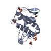
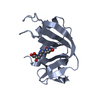
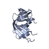
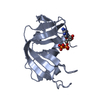
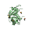
- Structure visualization
| Structure viewer | Molecule: MolmilJmol/JSmol |
|---|
- Downloads & links
-Download
| PDBx/mmCIF format | 5rsa.cif.gz | 64.2 KB | Display | PDBx/mmCIF format |
|---|---|---|---|---|
| PDB format | pdb5rsa.ent.gz | 49.7 KB | Display | PDB format |
| PDBx/mmJSON format | 5rsa.json.gz | Tree view | PDBx/mmJSON format | |
| Others |  Other downloads Other downloads |
-Validation report
| Arichive directory | https://data.pdbj.org/pub/pdb/validation_reports/rs/5rsaftp://data.pdbj.org/pub/pdb/validation_reports/rs/5rsa | HTTPS FTP |
|---|
-Related structure data
| Similar structure data |
|---|


-Links
 PDBj
PDBj


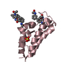
- Assembly
| Deposited unit | 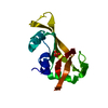
| ||||||||
|---|---|---|---|---|---|---|---|---|---|
| 1 |
| ||||||||
| Unit cell |
| ||||||||
| Atom site foot note | 1: RESIDUES 93 AND 114 ARE CIS-PROLINES. |
-Components
| #1: Protein | Mass: 13708.326 Da / Num. of mol.: 1 Source method: isolated from a genetically manipulated source Source: (gene. exp.) |
|---|---|
| #2: Chemical | ChemComp-PO4 /   Mass: 94.971 Da / Num. of mol.: 1 / Source method: obtained synthetically / Formula: PO4 Mass: 94.971 Da / Num. of mol.: 1 / Source method: obtained synthetically / Formula: PO4 |
| #3: Chemical | ChemComp-DOD /   Mass: 18.015 Da / Num. of mol.: 128 / Source method: isolated from a natural source / Formula: D2O Mass: 18.015 Da / Num. of mol.: 128 / Source method: isolated from a natural source / Formula: D2O |
| Has protein modification | Y |
-Experimental details
-Experiment
| Experiment |
|
|---|
- Sample preparation
| Crystal | Density Matthews: 2.17 Å3/Da / Density % sol: 43.25 % Description: DATA WERE COLLECTED ON DEUTERATED CRYSTALS. THE SOLVENT IS TERTIARY BUTANOL. AMIDE HYDROGENS WHICH EXCHANGED LESS THAN 50 PER CENT ARE ENTERED AS H, OTHERS ARE ENTERED AS D. | ||||||||||||||||||||||||
|---|---|---|---|---|---|---|---|---|---|---|---|---|---|---|---|---|---|---|---|---|---|---|---|---|---|
| Crystal grow | *PLUS pH: 5.3 / Method: unknown / Details: Wlodawer, A., (1982) J.Biol.Chem., 257, 1325. | ||||||||||||||||||||||||
| Components of the solutions | *PLUS
|
- Processing
| Refinement |
| ||||||||||||||||||||||||||||||||||||||||||||||||||||||||||||
|---|---|---|---|---|---|---|---|---|---|---|---|---|---|---|---|---|---|---|---|---|---|---|---|---|---|---|---|---|---|---|---|---|---|---|---|---|---|---|---|---|---|---|---|---|---|---|---|---|---|---|---|---|---|---|---|---|---|---|---|---|---|
| Refinement step | Cycle: LAST / Resolution: 2→10 Å
| ||||||||||||||||||||||||||||||||||||||||||||||||||||||||||||
| Refine LS restraints |
| ||||||||||||||||||||||||||||||||||||||||||||||||||||||||||||
| Refinement | *PLUS Num. reflection obs: 7708 / Rfactor obs: 0.159 / σ(I): 3 | ||||||||||||||||||||||||||||||||||||||||||||||||||||||||||||
| Solvent computation | *PLUS | ||||||||||||||||||||||||||||||||||||||||||||||||||||||||||||
| Displacement parameters | *PLUS | ||||||||||||||||||||||||||||||||||||||||||||||||||||||||||||
| Refine LS restraints | *PLUS
|
