 Movie
Movie Controller


+ Open data

- Basic information
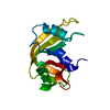
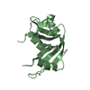
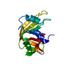
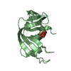
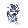
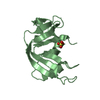
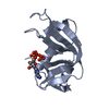
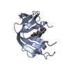
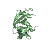
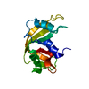
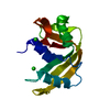
| Entry | Database: PDB / ID: 1jvu | ||||||
|---|---|---|---|---|---|---|---|
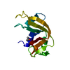
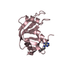
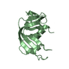
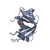
| Title | CRYSTAL STRUCTURE OF RIBONUCLEASE A (COMPLEXED FORM) | ||||||
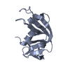
 Components Components | RIBONUCLEASE A | ||||||
 Keywords Keywords | HYDROLASE / PROTEIN DYNAMICS / PROTEIN STRUCTURE-FUNCTION | ||||||
| Function / homology |  Function and homology information Function and homology informationpancreatic ribonuclease / ribonuclease A activity / RNA nuclease activity / nucleic acid binding / defense response to Gram-positive bacterium / hydrolase activity / extracellular region Similarity search - Function | ||||||
| Biological species |  | ||||||
| Method |  X-RAY DIFFRACTION / MOLECULAR REPLACEMENT / Resolution: 1.78 Å X-RAY DIFFRACTION / MOLECULAR REPLACEMENT / Resolution: 1.78 Å | ||||||
 Authors Authors | Vitagliano, L. / Merlino, A. / Zagari, A. / Mazzarella, L. | ||||||
 Citation Citation | Journal: Proteins / Year: 2002 Title: Reversible Substrate-Induced Domain Motions in Ribonuclease A Authors: Vitagliano, L. / Merlino, A. / Zagari, A. / Mazzarella, L. #1: Journal: Protein Sci. / Year: 2000Title: Productive and Non-Productive Binding to Ribonuclease A: X-Ray Structure of Two Complexes with Uridylyl(2',5')Guanosine Authors: Vitagliano, L. / Merlino, A. / Zagari, A. / Mazzarella, L. #2: Journal: Protein Sci. / Year: 1998Title: Binding of a substrate analog to a domain swapping protein: X-ray structure of the complex of bovine seminal ribonuclease with uridylyl(2',5')adenosine Authors: Vitagliano, L. / Adinolfi, S. / Riccio, A. / Sica, F. / Zagari, A. / Mazzarella, L. | ||||||
| History |
|
- Structure visualization
| Structure viewer | Molecule: MolmilJmol/JSmol |
|---|
- Downloads & links
-Download
| PDBx/mmCIF format | 1jvu.cif.gz | 61.5 KB | Display | PDBx/mmCIF format |
|---|---|---|---|---|
| PDB format | pdb1jvu.ent.gz | 45 KB | Display | PDB format |
| PDBx/mmJSON format | 1jvu.json.gz | Tree view | PDBx/mmJSON format | |
| Others |  Other downloads Other downloads |
-Validation report
| Arichive directory | https://data.pdbj.org/pub/pdb/validation_reports/jv/1jvuftp://data.pdbj.org/pub/pdb/validation_reports/jv/1jvu | HTTPS FTP |
|---|
-Related structure data
| Related structure data |  1jvtC  1jvvC 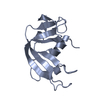 1afuS C: citing same article ( S: Starting model for refinement |
|---|---|
| Similar structure data |


-Links
 PDBj
PDBj

- Assembly
| Deposited unit | 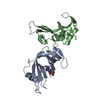
| ||||||||||
|---|---|---|---|---|---|---|---|---|---|---|---|
| 1 | 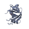
| ||||||||||
| 2 | 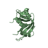
| ||||||||||
| Unit cell |
| ||||||||||
| Details | The biological assembly is a monomer |
-Components
| #1: Protein | Mass: 13708.326 Da / Num. of mol.: 2 / Source method: isolated from a natural source / Details: COMPLEXED WITH CYTIDINE-2'-MONOPHOSPHATE / Source: (natural) #2: Chemical | ChemComp-C2P / |   Mass: 323.197 Da / Num. of mol.: 1 / Source method: obtained synthetically / Formula: C9H14N3O8P Mass: 323.197 Da / Num. of mol.: 1 / Source method: obtained synthetically / Formula: C9H14N3O8P#3: Water | ChemComp-HOH / |  Mass: 18.015 Da / Num. of mol.: 84 / Source method: isolated from a natural source / Formula: H2O Mass: 18.015 Da / Num. of mol.: 84 / Source method: isolated from a natural source / Formula: H2OHas protein modification | Y | |
|---|
-Experimental details
-Experiment
| Experiment | Method: X-RAY DIFFRACTION / Number of used crystals: 1 |
|---|
- Sample preparation
| Crystal | Density Matthews: 2.26 Å3/Da / Density % sol: 45.46 % | ||||||||||||||||||||||||||||||||||||
|---|---|---|---|---|---|---|---|---|---|---|---|---|---|---|---|---|---|---|---|---|---|---|---|---|---|---|---|---|---|---|---|---|---|---|---|---|---|
| Crystal grow | Temperature: 298 K / Method: vapor diffusion, hanging drop / pH: 5 Details: PEG 4000, sodium citrate, pH 5.00, VAPOR DIFFUSION, HANGING DROP at 298K | ||||||||||||||||||||||||||||||||||||
| Crystal grow | *PLUS pH: 5 | ||||||||||||||||||||||||||||||||||||
| Components of the solutions | *PLUS
|
-Data collection
| Diffraction | Mean temperature: 298 K |
|---|---|
| Diffraction source | Source: ROTATING ANODE / Type: ENRAF-NONIUS FR591 / Wavelength: 1.5418 / Wavelength: 1.5418 Å |
| Detector | Type: MAC Science DIP-2030B / Detector: IMAGE PLATE / Date: Jul 30, 1999 |
| Radiation | Monochromator: DOUBLE MIRRORS / Protocol: SINGLE WAVELENGTH / Monochromatic (M) / Laue (L): M / Scattering type: x-ray |
| Radiation wavelength | Wavelength: 1.5418 Å / Relative weight: 1 |
| Reflection | Resolution: 1.78→20 Å / Num. all: 39019 / Num. obs: 22408 / % possible obs: 95 % / Observed criterion σ(F): 0 / Observed criterion σ(I): 0 / Redundancy: 1.7 % / Rmerge(I) obs: 0.053 |
| Reflection shell | Resolution: 1.78→1.86 Å / % possible all: 85 |
| Reflection | *PLUS Lowest resolution: 20 Å / % possible obs: 95 % / Num. measured all: 39019 |
- Processing
| Software |
| ||||||||||||||||||||||||||||||||||||||||||||||||||||||||||||
|---|---|---|---|---|---|---|---|---|---|---|---|---|---|---|---|---|---|---|---|---|---|---|---|---|---|---|---|---|---|---|---|---|---|---|---|---|---|---|---|---|---|---|---|---|---|---|---|---|---|---|---|---|---|---|---|---|---|---|---|---|---|
| Refinement | Method to determine structure: MOLECULAR REPLACEMENT Starting model: 1AFU Resolution: 1.78→8 Å / Isotropic thermal model: Isotropic / Cross valid method: THROUGHOUT / σ(F): 2 / Stereochemistry target values: Engh & Huber
| ||||||||||||||||||||||||||||||||||||||||||||||||||||||||||||
| Refinement step | Cycle: LAST / Resolution: 1.78→8 Å
| ||||||||||||||||||||||||||||||||||||||||||||||||||||||||||||
| Refine LS restraints |
| ||||||||||||||||||||||||||||||||||||||||||||||||||||||||||||
| LS refinement shell | Resolution: 1.78→1.86 Å
| ||||||||||||||||||||||||||||||||||||||||||||||||||||||||||||
| Refinement | *PLUS Lowest resolution: 8 Å | ||||||||||||||||||||||||||||||||||||||||||||||||||||||||||||
| Solvent computation | *PLUS | ||||||||||||||||||||||||||||||||||||||||||||||||||||||||||||
| Displacement parameters | *PLUS | ||||||||||||||||||||||||||||||||||||||||||||||||||||||||||||
| Refine LS restraints | *PLUS
| ||||||||||||||||||||||||||||||||||||||||||||||||||||||||||||
| LS refinement shell | *PLUS Rfactor Rfree: 0.33 / Rfactor Rwork: 0.28 / Rfactor obs: 0.28 |
