 Movie
Movie Controller


+ Open data

- Basic information
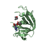
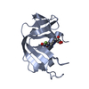
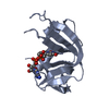
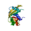
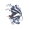
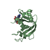
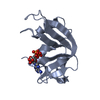
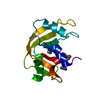
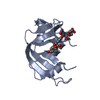
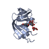
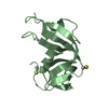
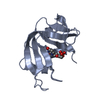
| Entry | Database: PDB / ID: 1o0f | ||||||
|---|---|---|---|---|---|---|---|
| Title | RNASE A in complex with 3',5'-ADP | ||||||
 Components Components | Ribonuclease pancreatic | ||||||
 Keywords Keywords | HYDROLASE / Ribonuclease | ||||||
| Function / homology |  Function and homology information Function and homology informationpancreatic ribonuclease / ribonuclease A activity / RNA nuclease activity / nucleic acid binding / defense response to Gram-positive bacterium / hydrolase activity / extracellular region Similarity search - Function | ||||||
| Biological species |  | ||||||
| Method |  X-RAY DIFFRACTION / FOURIER SYNTHESIS / Resolution: 1.5 Å X-RAY DIFFRACTION / FOURIER SYNTHESIS / Resolution: 1.5 Å | ||||||
 Authors Authors | Leonidas, D.D. / Oikonomakos, N.G. / Chrysina, E.D. / Kosmopoulou, M.N. / Vlassi, M. | ||||||
 Citation Citation | Journal: PROTEIN SCI. / Year: 2003 Title: High-resolution crystal structures of ribonuclease A complexed with adenylic and uridylic nucleotide inhibitors. Implications for structure-based design of ribonucleolytic inhibitors Authors: Leonidas, D.D. / Chavali, G.B. / Oikonomakos, N.G. / Chrysina, E.D. / Kosmopoulou, M.N. / Vlassi, M. / Frankling, C. / Acharya, K.R. | ||||||
| History |
|
- Structure visualization
| Structure viewer | Molecule: MolmilJmol/JSmol |
|---|
- Downloads & links
-Download
| PDBx/mmCIF format | 1o0f.cif.gz | 71.3 KB | Display | PDBx/mmCIF format |
|---|---|---|---|---|
| PDB format | pdb1o0f.ent.gz | 52.3 KB | Display | PDB format |
| PDBx/mmJSON format | 1o0f.json.gz | Tree view | PDBx/mmJSON format | |
| Others |  Other downloads Other downloads |
-Validation report
| Arichive directory | https://data.pdbj.org/pub/pdb/validation_reports/o0/1o0fftp://data.pdbj.org/pub/pdb/validation_reports/o0/1o0f | HTTPS FTP |
|---|
-Related structure data
| Related structure data | 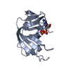 1o0hC 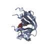 1o0mC 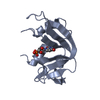 1o0nC 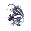 1o0oC 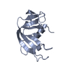 1afuS C: citing same article ( S: Starting model for refinement |
|---|---|
| Similar structure data |

-Links
 PDBj
PDBj


- Assembly
| Deposited unit | 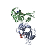
| |||||||||||||||
|---|---|---|---|---|---|---|---|---|---|---|---|---|---|---|---|---|
| 1 | 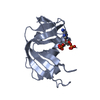
| |||||||||||||||
| 2 | 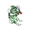
| |||||||||||||||
| Unit cell |
| |||||||||||||||
| Components on special symmetry positions |
|
-Components
| #1: Protein | Mass: 13708.326 Da / Num. of mol.: 2 / Source method: isolated from a natural source / Source: (natural) #2: Chemical | 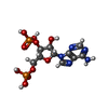  Type: RNA linking / Mass: 427.201 Da / Num. of mol.: 2 / Source method: obtained synthetically / Formula: C10H15N5O10P2 Type: RNA linking / Mass: 427.201 Da / Num. of mol.: 2 / Source method: obtained synthetically / Formula: C10H15N5O10P2#3: Water | ChemComp-HOH / |  Mass: 18.015 Da / Num. of mol.: 389 / Source method: isolated from a natural source / Formula: H2O Mass: 18.015 Da / Num. of mol.: 389 / Source method: isolated from a natural source / Formula: H2OHas protein modification | Y | |
|---|
-Experimental details
-Experiment
| Experiment | Method: X-RAY DIFFRACTION / Number of used crystals: 1 |
|---|
- Sample preparation
| Crystal | Density Matthews: 1.79 Å3/Da / Density % sol: 30.68 % | ||||||||||||||||||||||||||||||||||||
|---|---|---|---|---|---|---|---|---|---|---|---|---|---|---|---|---|---|---|---|---|---|---|---|---|---|---|---|---|---|---|---|---|---|---|---|---|---|
| Crystal grow | Temperature: 289 K / Method: vapor diffusion, hanging drop / pH: 5.5 Details: 20% PEG 4000, 20MM SODIUM CITRATE, pH 5.5, VAPOR DIFFUSION, HANGING DROP, temperature 289K | ||||||||||||||||||||||||||||||||||||
| Crystal grow | *PLUS Temperature: 16 ℃ / Method: vapor diffusion, hanging drop / Details: Leonidas, D.D., (1997) Biochemistry, 36, 5578. | ||||||||||||||||||||||||||||||||||||
| Components of the solutions | *PLUS
|
-Data collection
| Diffraction | Mean temperature: 100 K |
|---|---|
| Diffraction source | Source: ROTATING ANODE / Type: RIGAKU / Wavelength: 1.5419 Å |
| Detector | Type: RIGAKU RAXIS IV / Detector: IMAGE PLATE / Date: Nov 25, 1999 / Details: mirrors |
| Radiation | Monochromator: MIRROR + Ni FILTER / Protocol: SINGLE WAVELENGTH / Monochromatic (M) / Laue (L): M / Scattering type: x-ray |
| Radiation wavelength | Wavelength: 1.5419 Å / Relative weight: 1 |
| Reflection | Resolution: 1.5→30 Å / Num. all: 36948 / Num. obs: 36948 / % possible obs: 97.9 % / Observed criterion σ(F): 1 / Observed criterion σ(I): -3 / Redundancy: 11.5 % / Biso Wilson estimate: 23.3 Å2 / Rsym value: 0.062 / Net I/σ(I): 14.9 |
| Reflection shell | Resolution: 1.5→1.55 Å / Rsym value: 0.369 / % possible all: 95.5 |
| Reflection | *PLUS Num. measured all: 423949 / Rmerge(I) obs: 0.062 |
| Reflection shell | *PLUS Highest resolution: 1.5 Å / % possible obs: 95.5 % / Rmerge(I) obs: 0.369 / Mean I/σ(I) obs: 3.6 |
- Processing
| Software |
| ||||||||||||||||||||||||||||||||||||
|---|---|---|---|---|---|---|---|---|---|---|---|---|---|---|---|---|---|---|---|---|---|---|---|---|---|---|---|---|---|---|---|---|---|---|---|---|---|
| Refinement | Method to determine structure: FOURIER SYNTHESIS Starting model: PDB ENTRY 1AFU Resolution: 1.5→29.02 Å / Rfactor Rfree error: 0.006 / Data cutoff high absF: 958585.22 / Data cutoff low absF: 0 / Isotropic thermal model: RESTRAINED / Cross valid method: THROUGHOUT / σ(F): 0.1 / Stereochemistry target values: Engh & Huber
| ||||||||||||||||||||||||||||||||||||
| Solvent computation | Solvent model: FLAT MODEL / Bsol: 36.8564 Å2 / ksol: 0.361218 e/Å3 | ||||||||||||||||||||||||||||||||||||
| Displacement parameters | Biso mean: 23.4 Å2
| ||||||||||||||||||||||||||||||||||||
| Refine analyze |
| ||||||||||||||||||||||||||||||||||||
| Refinement step | Cycle: LAST / Resolution: 1.5→29.02 Å
| ||||||||||||||||||||||||||||||||||||
| Refine LS restraints |
| ||||||||||||||||||||||||||||||||||||
| LS refinement shell | Resolution: 1.5→1.59 Å / Rfactor Rfree error: 0.026 / Total num. of bins used: 6
| ||||||||||||||||||||||||||||||||||||
| Xplor file |
| ||||||||||||||||||||||||||||||||||||
| Refinement | *PLUS Highest resolution: 1.5 Å / % reflection Rfree: 5 % | ||||||||||||||||||||||||||||||||||||
| Solvent computation | *PLUS | ||||||||||||||||||||||||||||||||||||
| Displacement parameters | *PLUS | ||||||||||||||||||||||||||||||||||||
| Refine LS restraints | *PLUS
| ||||||||||||||||||||||||||||||||||||
| LS refinement shell | *PLUS Rfactor Rfree: 0.452 / Rfactor Rwork: 0.446 |
