 Movie
Movie Controller

[English] 日本語
 Yorodumi
- PDB-1f0v: Crystal structure of an Rnase A dimer displaying a new type of 3D... -
+ Open data

- Basic information
| Entry | Database: PDB / ID: 1f0v | ||||||
|---|---|---|---|---|---|---|---|
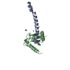
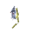
| Title | Crystal structure of an Rnase A dimer displaying a new type of 3D domain swapping | ||||||
 Components Components |
| ||||||
 Keywords Keywords | hydrolase/DNA / domain swapping / crystal / ribonuclease / bovine pancreas / hydrolase-DNA COMPLEX | ||||||
| Function / homology |  Function and homology information Function and homology informationpancreatic ribonuclease / ribonuclease A activity / RNA nuclease activity / nucleic acid binding / defense response to Gram-positive bacterium / hydrolase activity / extracellular region Similarity search - Function | ||||||
| Biological species |  | ||||||
| Method |  X-RAY DIFFRACTION / SYNCHROTRON / Resolution: 1.7 Å X-RAY DIFFRACTION / SYNCHROTRON / Resolution: 1.7 Å | ||||||
 Authors Authors | Liu, Y.S. / Gotte, G. / Libonati, M. / Eisenberg, D.S. | ||||||
 Citation Citation | Journal: Nat.Struct.Biol. / Year: 2001 Title: A domain-swapped RNase A dimer with implications for amyloid formation Authors: Liu, Y.S. / Gotte, G. / Libonati, M. / Eisenberg, D.S. | ||||||
| History |
|
- Structure visualization
| Structure viewer | Molecule: MolmilJmol/JSmol |
|---|
- Downloads & links
-Download
| PDBx/mmCIF format | 1f0v.cif.gz | 131.8 KB | Display | PDBx/mmCIF format |
|---|---|---|---|---|
| PDB format | pdb1f0v.ent.gz | 101.4 KB | Display | PDB format |
| PDBx/mmJSON format | 1f0v.json.gz | Tree view | PDBx/mmJSON format | |
| Others |  Other downloads Other downloads |
-Validation report
| Arichive directory | https://data.pdbj.org/pub/pdb/validation_reports/f0/1f0vftp://data.pdbj.org/pub/pdb/validation_reports/f0/1f0v | HTTPS FTP |
|---|
-Related structure data
| Related structure data | |
|---|---|
| Similar structure data |


-Links
 PDBj
PDBj

- Assembly
| Deposited unit | 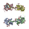
| ||||||||||
|---|---|---|---|---|---|---|---|---|---|---|---|
| 1 | 
| ||||||||||
| 2 | 
| ||||||||||
| 3 | 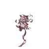
| ||||||||||
| 4 | 
| ||||||||||
| Unit cell |
|
-Components
| #1: DNA chain | Mass: 573.430 Da / Num. of mol.: 4 / Source method: obtained synthetically #2: Protein | Mass: 13708.326 Da / Num. of mol.: 4 / Source method: isolated from a natural source / Source: (natural) #3: Chemical | ChemComp-PO4 /   Mass: 94.971 Da / Num. of mol.: 4 / Source method: obtained synthetically / Formula: PO4 Mass: 94.971 Da / Num. of mol.: 4 / Source method: obtained synthetically / Formula: PO4#4: Chemical | ChemComp-GOL /   Mass: 92.094 Da / Num. of mol.: 24 / Source method: obtained synthetically / Formula: C3H8O3 Mass: 92.094 Da / Num. of mol.: 24 / Source method: obtained synthetically / Formula: C3H8O3#5: Water | ChemComp-HOH / |  Mass: 18.015 Da / Num. of mol.: 554 / Source method: isolated from a natural source / Formula: H2O Mass: 18.015 Da / Num. of mol.: 554 / Source method: isolated from a natural source / Formula: H2OHas protein modification | Y | |
|---|
-Experimental details
-Experiment
| Experiment | Method: X-RAY DIFFRACTION / Number of used crystals: 1 |
|---|
- Sample preparation
| Crystal | Density Matthews: 3.23 Å3/Da / Density % sol: 61.96 % | |||||||||||||||||||||||||||||||||||
|---|---|---|---|---|---|---|---|---|---|---|---|---|---|---|---|---|---|---|---|---|---|---|---|---|---|---|---|---|---|---|---|---|---|---|---|---|
| Crystal grow | Temperature: 293 K / Method: vapor diffusion, hanging drop / pH: 6.5 Details: 0.1 M Phosphate buffer, 16 % PEG 4000, 2 % Dioxane, pH 6.5, VAPOR DIFFUSION, HANGING DROP, temperature 293K | |||||||||||||||||||||||||||||||||||
| Components of the solutions |
| |||||||||||||||||||||||||||||||||||
| Crystal grow | *PLUS Details: drop contains protein and reservoir solution in a 1:1 ratio | |||||||||||||||||||||||||||||||||||
| Components of the solutions | *PLUS
|
-Data collection
| Diffraction | Mean temperature: 100 K |
|---|---|
| Diffraction source | Source: SYNCHROTRON / Site: NSLS  / Beamline: X12B / Wavelength: 1.072 / Beamline: X12B / Wavelength: 1.072 |
| Detector | Detector: AREA DETECTOR |
| Radiation | Protocol: SINGLE WAVELENGTH / Monochromatic (M) / Laue (L): M / Scattering type: x-ray |
| Radiation wavelength | Wavelength: 1.072 Å / Relative weight: 1 |
| Reflection | Resolution: 1.7→100 Å / Num. all: 76047 / Num. obs: 76047 / % possible obs: 95 % / Observed criterion σ(F): 0 / Observed criterion σ(I): 0 / Redundancy: 10 % / Biso Wilson estimate: 20 Å2 / Rmerge(I) obs: 0.052 / Net I/σ(I): 14 |
| Reflection shell | Resolution: 1.7→1.76 Å / Redundancy: 3 % / Rmerge(I) obs: 0.08 / Num. unique all: 7548 / % possible all: 95 |
| Reflection | *PLUS Lowest resolution: 40 Å / Num. measured all: 813673 |
| Reflection shell | *PLUS % possible obs: 95 % |
- Processing
| Software |
| |||||||||||||||||||||||||
|---|---|---|---|---|---|---|---|---|---|---|---|---|---|---|---|---|---|---|---|---|---|---|---|---|---|---|
| Refinement | Resolution: 1.7→10 Å / Cross valid method: THROUGHOUT / σ(F): 0 / σ(I): 0 / Stereochemistry target values: Engh & Huber
| |||||||||||||||||||||||||
| Refinement step | Cycle: LAST / Resolution: 1.7→10 Å
| |||||||||||||||||||||||||
| Refine LS restraints |
| |||||||||||||||||||||||||
| Software | *PLUS Name: CNS / Classification: refinement | |||||||||||||||||||||||||
| Refinement | *PLUS Num. reflection all: 75824 | |||||||||||||||||||||||||
| Solvent computation | *PLUS | |||||||||||||||||||||||||
| Displacement parameters | *PLUS Biso mean: 25.14 Å2 |
