 Movie
Movie Controller

[English] 日本語
 Yorodumi
- PDB-1rsm: THE 2-ANGSTROMS RESOLUTION STRUCTURE OF A THERMOSTABLE RIBONUCLEA... -
+ Open data

- Basic information
| Entry | Database: PDB / ID: 1rsm | ||||||
|---|---|---|---|---|---|---|---|
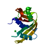
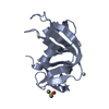
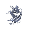
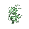
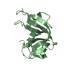
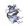
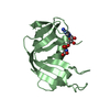
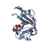
| Title | THE 2-ANGSTROMS RESOLUTION STRUCTURE OF A THERMOSTABLE RIBONUCLEASE A CHEMICALLY CROSS-LINKED BETWEEN LYSINE RESIDUES 7 AND 41 | ||||||
 Components Components | RIBONUCLEASE A | ||||||
 Keywords Keywords | HYDROLASE (NUCLEIC ACID / RNA) | ||||||
| Function / homology |  Function and homology information Function and homology informationpancreatic ribonuclease / ribonuclease A activity / RNA nuclease activity / nucleic acid binding / defense response to Gram-positive bacterium / hydrolase activity / extracellular region Similarity search - Function | ||||||
| Biological species |  | ||||||
| Method |  X-RAY DIFFRACTION / Resolution: 2 Å X-RAY DIFFRACTION / Resolution: 2 Å | ||||||
 Authors Authors | Weber, P.C. / Sheriff, S. / Ohlendorf, D.H. / Finzel, B.C. / Salemme, F.R. | ||||||
 Citation Citation | Journal: Proc.Natl.Acad.Sci.USA / Year: 1985 Title: The 2-A resolution structure of a thermostable ribonuclease A chemically cross-linked between lysine residues 7 and 41. Authors: Weber, P.C. / Sheriff, S. / Ohlendorf, D.H. / Finzel, B.C. / Salemme, F.R. #1: Journal: J.Mol.Biol. / Year: 1985Title: Preliminary Crystallographic Data for Cross-Linked (Lysine7-Lysine41)-Ribonuclease A Authors: Weber, P.C. / Salemme, F.R. / Lin, S.H. / Konishi, Y. / Scheraga, H.A. #2: Journal: Biochemistry / Year: 1984Title: Influence of an Extrinsic Cross-Link on the Folding Pathway of Ribonuclease A. Conformational and Thermodynamic Analysis of Cross-Linked (Lysine7-Lysine41)-Ribonuclease A Authors: Lin, S.H. / Konishi, Y. / Denton, M.E. / Scheraga, H.A. #3: Journal: Biochemistry / Year: 1983Title: Structure of Ribonuclease A. Results of Joint Neutron and X-Ray Refinement at 2.0-Angstroms Resolutions Authors: Wlodawer, A. / Sjolin, L. | ||||||
| History |
| ||||||
| Remark 700 | SHEET THIS STRUCTURE CONTAINS TWO SHEETS. SHEET S1 COMPRISES THREE STRANDS. IN THE SECOND STRAND OF ...SHEET THIS STRUCTURE CONTAINS TWO SHEETS. SHEET S1 COMPRISES THREE STRANDS. IN THE SECOND STRAND OF SHEET S1, RESIDUES 88 AND 89 *BULGE OUT*. IN ORDER TO REPRESENT THIS BREAK IN STRAND 2, TWO SHEETS (S1A AND S1B) ARE DEFINED BELOW. STRANDS 1 AND 3 OF *SHEETS* S1A AND S1B ARE, THEREFORE, IDENTICAL AND STRAND 2 DIFFERS. SHEET S2 COMPRISES FOUR STRANDS. RESIDUE 120 DOES NOT PROPERLY BELONG IN STRAND 4 OF SHEET S2. IN ORDER TO REPRESENT THIS BREAK IN STRAND 4, TWO SHEETS (S2A AND S2B) ARE DEFINED BELOW. STRANDS 1,2,3 OF *SHEETS* S2A AND S2B ARE, THEREFORE, IDENTICAL AND STRAND 4 DIFFERS. |
- Structure visualization
| Structure viewer | Molecule: MolmilJmol/JSmol |
|---|
- Downloads & links
-Download
| PDBx/mmCIF format | 1rsm.cif.gz | 38.1 KB | Display | PDBx/mmCIF format |
|---|---|---|---|---|
| PDB format | pdb1rsm.ent.gz | 26 KB | Display | PDB format |
| PDBx/mmJSON format | 1rsm.json.gz | Tree view | PDBx/mmJSON format | |
| Others |  Other downloads Other downloads |
-Validation report
| Arichive directory | https://data.pdbj.org/pub/pdb/validation_reports/rs/1rsmftp://data.pdbj.org/pub/pdb/validation_reports/rs/1rsm | HTTPS FTP |
|---|
-Related structure data
| Similar structure data |
|---|


-Links
 PDBj
PDBj

- Assembly
| Deposited unit | 
| ||||||||
|---|---|---|---|---|---|---|---|---|---|
| 1 |
| ||||||||
| Unit cell |
| ||||||||
| Atom site foot note | 1: RESIDUES 93 AND 114 ARE CIS-PROLINES. |
-Components
| #1: Protein | Mass: 13708.326 Da / Num. of mol.: 1 Source method: isolated from a genetically manipulated source Source: (gene. exp.) |
|---|---|
| #2: Chemical | ChemComp-NIN /   Mass: 168.107 Da / Num. of mol.: 1 / Source method: obtained synthetically / Formula: C6H4N2O4 Mass: 168.107 Da / Num. of mol.: 1 / Source method: obtained synthetically / Formula: C6H4N2O4 |
| #3: Water | ChemComp-HOH /  Mass: 18.015 Da / Num. of mol.: 75 / Source method: isolated from a natural source / Formula: H2O Mass: 18.015 Da / Num. of mol.: 75 / Source method: isolated from a natural source / Formula: H2O |
| Has protein modification | Y |
-Experimental details
-Experiment
| Experiment | Method: X-RAY DIFFRACTION |
|---|
- Sample preparation
| Crystal | Density Matthews: 2.11 Å3/Da / Density % sol: 41.65 % |
|---|---|
| Crystal grow | *PLUS Temperature: 7 ℃ / pH: 8 / Method: microdialysis |
| Components of the solutions | *PLUS Conc.: 30 %(v/v) / Common name: ethanolDetails: 50mM-Tris-HCl(pH7.5) and 50 mM-imidazole(pH8.0) are also suitable |
-Data collection
| Radiation | Scattering type: x-ray |
|---|---|
| Radiation wavelength | Relative weight: 1 |
| Reflection | *PLUS Highest resolution: 2 Å / Lowest resolution: 2.1 Å / Num. obs: 8301 / Rmerge(I) obs: 0.081 / Num. measured all: 10701 |
- Processing
| Software | Name: PROLSQ / Classification: refinement | ||||||||||||||||||||||||||||||||||||||||||||||||||||||||||||
|---|---|---|---|---|---|---|---|---|---|---|---|---|---|---|---|---|---|---|---|---|---|---|---|---|---|---|---|---|---|---|---|---|---|---|---|---|---|---|---|---|---|---|---|---|---|---|---|---|---|---|---|---|---|---|---|---|---|---|---|---|---|
| Refinement | Highest resolution: 2 Å / σ(I): 1 /
| ||||||||||||||||||||||||||||||||||||||||||||||||||||||||||||
| Refinement step | Cycle: LAST / Highest resolution: 2 Å
| ||||||||||||||||||||||||||||||||||||||||||||||||||||||||||||
| Refine LS restraints |
| ||||||||||||||||||||||||||||||||||||||||||||||||||||||||||||
| Refinement | *PLUS Lowest resolution: 20 Å / σ(I): 1 / Highest resolution: 2 Å / Num. reflection obs: 7853 / Rfactor obs: 0.184 | ||||||||||||||||||||||||||||||||||||||||||||||||||||||||||||
| Solvent computation | *PLUS | ||||||||||||||||||||||||||||||||||||||||||||||||||||||||||||
| Displacement parameters | *PLUS | ||||||||||||||||||||||||||||||||||||||||||||||||||||||||||||
| Refine LS restraints | *PLUS
|
