 Movie
Movie Controller

[English] 日本語
 Yorodumi
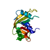
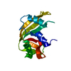
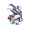
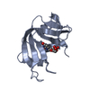
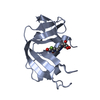
- PDB-3rn3: SEGMENTED ANISOTROPIC REFINEMENT OF BOVINE RIBONUCLEASE A BY THE ... -
+ Open data

- Basic information
| Entry | Database: PDB / ID: 3rn3 | |||||||||
|---|---|---|---|---|---|---|---|---|---|---|
| Title | SEGMENTED ANISOTROPIC REFINEMENT OF BOVINE RIBONUCLEASE A BY THE APPLICATION OF THE RIGID-BODY TLS MODEL | |||||||||
 Components Components | RIBONUCLEASE A | |||||||||
 Keywords Keywords | HYDROLASE (NUCLEIC ACID / RNA) | |||||||||
| Function / homology |  Function and homology information Function and homology informationpancreatic ribonuclease / ribonuclease A activity / RNA nuclease activity / nucleic acid binding / defense response to Gram-positive bacterium / hydrolase activity / extracellular region Similarity search - Function | |||||||||
| Biological species |  | |||||||||
| Method |  X-RAY DIFFRACTION / Resolution: 1.45 Å X-RAY DIFFRACTION / Resolution: 1.45 Å | |||||||||
 Authors Authors | Howlin, B. / Moss, D.S. / Harris, G.W. / Palmer, R.A. | |||||||||
 Citation Citation | Journal: Acta Crystallogr.,Sect.A / Year: 1989 Title: Segmented anisotropic refinement of bovine ribonuclease A by the application of the rigid-body TLS model. Authors: Howlin, B. / Moss, D.S. / Harris, G.W. #1: Journal: Biochim.Biophys.Acta / Year: 1987Title: Ribonuclease A. Analysis of the Hydrogen Bond Geometry, and Spatial Accessibility at the Active Site Authors: Harris, G.W. / Borkakoti, N. / Moss, D.S. / Palmer, R.A. / Howlin, B. #2: Journal: Acta Crystallogr.,Sect.B / Year: 1986Title: Comparison of Two Independently Refined Models of Ribonuclease-A Authors: Wlodawer, A. / Borkakoti, N. / Moss, D.S. / Howlin, B. #3: Journal: J.Crystallogr.Spectrosc.Res. / Year: 1984Title: The Refined Structure of Ribonuclease-A at 1.45 Angstroms Resolution Authors: Borkakoti, N. / Moss, D.S. / Stanford, M.J. / Palmer, R.A. #4: Journal: J.Mol.Biol. / Year: 1983Title: Specificity of Pancreatic Ribonuclease-A. An X-Ray Study of a Protein-Nucleotide Complex Authors: Borkakoti, N. / Palmer, R.A. / Haneef, I. / Moss, D.S. #5: Journal: Eur.J.Biochem. / Year: 1983Title: The Active Site of Ribonuclease A from the Crystallographic Studies of Ribonuclease-A-Inhibitor Complexes Authors: Borkakoti, N. #6: Journal: Acta Crystallogr.,Sect.B / Year: 1982Title: Ribonuclease-A. Least-Squares Refinement of the Structure at 1.45 Angstroms Resolution Authors: Borkakoti, N. / Moss, D.S. / Palmer, R.A. #7: Journal: J.Mol.Biol. / Year: 1974Title: The Structure of Ribonuclease At 2.5 Angstrom Resolution Authors: Carlisle, C.H. / Palmer, R.A. / Mazumdar, S.K. / Gorinsky, B.A. / Yeates, D.G.R. | |||||||||
| History |
|
- Structure visualization
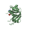
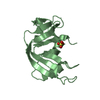
| Structure viewer | Molecule: MolmilJmol/JSmol |
|---|
- Downloads & links
-Download
| PDBx/mmCIF format | 3rn3.cif.gz | 37.4 KB | Display | PDBx/mmCIF format |
|---|---|---|---|---|
| PDB format | pdb3rn3.ent.gz | 25.2 KB | Display | PDB format |
| PDBx/mmJSON format | 3rn3.json.gz | Tree view | PDBx/mmJSON format | |
| Others |  Other downloads Other downloads |
-Validation report
| Arichive directory | https://data.pdbj.org/pub/pdb/validation_reports/rn/3rn3ftp://data.pdbj.org/pub/pdb/validation_reports/rn/3rn3 | HTTPS FTP |
|---|
-Related structure data
| Similar structure data |
|---|


-Links
 PDBj
PDBj

- Assembly
| Deposited unit | 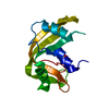
| ||||||||
|---|---|---|---|---|---|---|---|---|---|
| 1 |
| ||||||||
| Unit cell |
| ||||||||
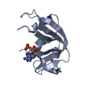
| Atom site foot note | 1: RESIDUES 93 AND 114 ARE CIS-PROLINES. / 2: SEE REMARK 5. |
-Components
| #1: Protein | Mass: 13708.326 Da / Num. of mol.: 1 Source method: isolated from a genetically manipulated source Source: (gene. exp.) |
|---|---|
| #2: Chemical | ChemComp-SO4 /   Mass: 96.063 Da / Num. of mol.: 1 / Source method: obtained synthetically / Formula: SO4 Mass: 96.063 Da / Num. of mol.: 1 / Source method: obtained synthetically / Formula: SO4 |
| #3: Water | ChemComp-HOH /  Mass: 18.015 Da / Num. of mol.: 107 / Source method: isolated from a natural source / Formula: H2O Mass: 18.015 Da / Num. of mol.: 107 / Source method: isolated from a natural source / Formula: H2O |
| Compound details | A SECOND SITE HAS BEEN LOCATED AND REFINED FOR HIS 119. |
| Has protein modification | Y |
-Experimental details
-Experiment
| Experiment | Method: X-RAY DIFFRACTION |
|---|
- Sample preparation
| Crystal | Density Matthews: 2.18 Å3/Da / Density % sol: 43.57 % |
|---|
- Processing
| Software | Name: RESTRAIN / Classification: refinement | |||||||||||||||||||||||||||||||||||||||||||||||||||||||||||||||
|---|---|---|---|---|---|---|---|---|---|---|---|---|---|---|---|---|---|---|---|---|---|---|---|---|---|---|---|---|---|---|---|---|---|---|---|---|---|---|---|---|---|---|---|---|---|---|---|---|---|---|---|---|---|---|---|---|---|---|---|---|---|---|---|---|
| Refinement | Highest resolution: 1.45 Å Details: THE CARBOXAMIDE TERMINI OF RESIDUES ASN 24, ASN 27, ASN 67, GLN 69, ASN 71, GLN 74, ASN 94, AND ASN 103 WERE "FLIPPED" BY 180 DEGREES TO CONFORM TO THE ASSIGNMENT OBSERVED IN THE STRUCTURE ...Details: THE CARBOXAMIDE TERMINI OF RESIDUES ASN 24, ASN 27, ASN 67, GLN 69, ASN 71, GLN 74, ASN 94, AND ASN 103 WERE "FLIPPED" BY 180 DEGREES TO CONFORM TO THE ASSIGNMENT OBSERVED IN THE STRUCTURE DEPOSITED BY WLODAWER ET AL (ENTRY 7RSA).
| |||||||||||||||||||||||||||||||||||||||||||||||||||||||||||||||
| Refinement step | Cycle: LAST / Highest resolution: 1.45 Å
| |||||||||||||||||||||||||||||||||||||||||||||||||||||||||||||||
| Refine LS restraints |
| |||||||||||||||||||||||||||||||||||||||||||||||||||||||||||||||
| Refinement | *PLUS Highest resolution: 1.45 Å / Num. reflection obs: 19238 / Rfactor obs: 0.2233 | |||||||||||||||||||||||||||||||||||||||||||||||||||||||||||||||
| Solvent computation | *PLUS | |||||||||||||||||||||||||||||||||||||||||||||||||||||||||||||||
| Displacement parameters | *PLUS |
