 Movie
Movie Controller

[English] 日本語
 Yorodumi
- PDB-1bzq: COMPLEX OF A DROMEDARY SINGLE-DOMAIN VHH ANTIBODY FRAGMENT WITH R... -
+ Open data

- Basic information
| Entry | Database: PDB / ID: 1bzq | ||||||
|---|---|---|---|---|---|---|---|
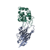
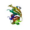
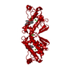
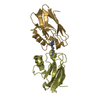
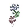
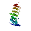
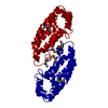
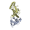
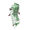
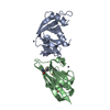
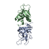
| Title | COMPLEX OF A DROMEDARY SINGLE-DOMAIN VHH ANTIBODY FRAGMENT WITH RNASE A | ||||||
 Components Components |
| ||||||
 Keywords Keywords | HYDROLASE/IMMUNE SYSTEM / IMMUNOGLUBULIN / ANTIBODY COMPLEX / RIBONUCLEASE / HYDROLASE-IMMUNE SYSTEM COMPLEX | ||||||
| Function / homology |  Function and homology information Function and homology informationpancreatic ribonuclease / ribonuclease A activity / RNA nuclease activity / nucleic acid binding / defense response to Gram-positive bacterium / hydrolase activity / extracellular region Similarity search - Function | ||||||
| Biological species |  | ||||||
| Method |  X-RAY DIFFRACTION / MOLECULAR REPLACEMENT / Resolution: 2.8 Å X-RAY DIFFRACTION / MOLECULAR REPLACEMENT / Resolution: 2.8 Å | ||||||
 Authors Authors | Decanniere, K. / Desmyter, A. / Gahroudhi, M. / Lauwereys, M. / Muyldermans, S. / Wyns, L. | ||||||
 Citation Citation | Journal: Structure Fold.Des. / Year: 1999 Title: A single-domain antibody fragment in complex with RNase A: non-canonical loop structures and nanomolar affinity using two CDR loops. Authors: Decanniere, K. / Desmyter, A. / Lauwereys, M. / Ghahroudi, M.A. / Muyldermans, S. / Wyns, L. #1: Journal: Nat.Struct.Biol. / Year: 1998Title: Crystal Structure of a Camel Single-Domain VH Antibody Fragment in Complex with Lysozyme Authors: Desmyter, A. / Transue, T.R. / Ghahroudi, M.A. / Thi, M.H. / Poortmans, F. / Hmaers, R. / Muyldermans, S. / WYNS, L. | ||||||
| History |
|
- Structure visualization
| Structure viewer | Molecule: MolmilJmol/JSmol |
|---|
- Downloads & links
-Download
| PDBx/mmCIF format | 1bzq.cif.gz | 193.6 KB | Display | PDBx/mmCIF format |
|---|---|---|---|---|
| PDB format | pdb1bzq.ent.gz | 154.5 KB | Display | PDB format |
| PDBx/mmJSON format | 1bzq.json.gz | Tree view | PDBx/mmJSON format | |
| Others |  Other downloads Other downloads |
-Validation report
| Arichive directory | https://data.pdbj.org/pub/pdb/validation_reports/bz/1bzqftp://data.pdbj.org/pub/pdb/validation_reports/bz/1bzq | HTTPS FTP |
|---|
-Related structure data

-Links
 PDBj
PDBj


- Assembly
| Deposited unit | 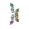
| ||||||||||||||||||||||||||||
|---|---|---|---|---|---|---|---|---|---|---|---|---|---|---|---|---|---|---|---|---|---|---|---|---|---|---|---|---|---|
| 1 | 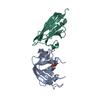
| ||||||||||||||||||||||||||||
| 2 | 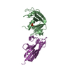
| ||||||||||||||||||||||||||||
| 3 | 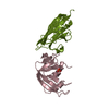
| ||||||||||||||||||||||||||||
| 4 | 
| ||||||||||||||||||||||||||||
| Unit cell |
| ||||||||||||||||||||||||||||
| Noncrystallographic symmetry (NCS) | NCS oper:
|
-Components
| #1: Protein | Mass: 13708.326 Da / Num. of mol.: 4 / Source method: isolated from a natural source / Details: RNASE A SUPPLIED BY SIGMA / Source: (natural) #2: Antibody | Mass: 13331.775 Da / Num. of mol.: 4 / Fragment: VARIABLE DOMAIN Source method: isolated from a genetically manipulated source Source: (gene. exp.)  #3: Chemical | ChemComp-PO4 /   Mass: 94.971 Da / Num. of mol.: 4 / Source method: obtained synthetically / Formula: PO4 Mass: 94.971 Da / Num. of mol.: 4 / Source method: obtained synthetically / Formula: PO4Has protein modification | Y | |
|---|
-Experimental details
-Experiment
| Experiment | Method: X-RAY DIFFRACTION / Number of used crystals: 2 |
|---|
- Sample preparation
| Crystal | Density Matthews: 2.11 Å3/Da / Density % sol: 50.77 % | ||||||||||||||||||||
|---|---|---|---|---|---|---|---|---|---|---|---|---|---|---|---|---|---|---|---|---|---|
| Crystal grow | Method: vapor diffusion, hanging drop / pH: 6.4 Details: HANGING DROP METHOD. 5 MG/ML OF THE COMPLEX RESERVOIR SOLUTION IS 0.1 M POTASSIUM PHOSPHATE PH 6.4 15 TO 20% W/V PEG 8000, vapor diffusion - hanging drop | ||||||||||||||||||||
| Crystal grow | *PLUS | ||||||||||||||||||||
| Components of the solutions | *PLUS
|
-Data collection
| Diffraction | Mean temperature: 293 K |
|---|---|
| Diffraction source | Source: ROTATING ANODE / Type: RIGAKU / Wavelength: 1.5418 |
| Detector | Type: MARRESEARCH / Detector: IMAGE PLATE |
| Radiation | Monochromator: SI CRYSTAL / Protocol: SINGLE WAVELENGTH / Monochromatic (M) / Laue (L): M / Scattering type: x-ray |
| Radiation wavelength | Wavelength: 1.5418 Å / Relative weight: 1 |
| Reflection | Resolution: 2.8→15 Å / Num. obs: 25413 / % possible obs: 99.2 % / Redundancy: 3.8 % / Rsym value: 0.111 / Net I/σ(I): 8.5 |
| Reflection shell | Resolution: 2.8→2.9 Å / Redundancy: 3.9 % / Mean I/σ(I) obs: 3.9 / Rsym value: 0.336 / % possible all: 98.2 |
| Reflection | *PLUS Rmerge(I) obs: 0.111 |
| Reflection shell | *PLUS % possible obs: 98.2 % / Rmerge(I) obs: 0.336 / Mean I/σ(I) obs: 3.7 |
- Processing
| Software |
| ||||||||||||||||||||||||||||||||||||||||||||||||||||||||||||||||||||||||||||||||||||
|---|---|---|---|---|---|---|---|---|---|---|---|---|---|---|---|---|---|---|---|---|---|---|---|---|---|---|---|---|---|---|---|---|---|---|---|---|---|---|---|---|---|---|---|---|---|---|---|---|---|---|---|---|---|---|---|---|---|---|---|---|---|---|---|---|---|---|---|---|---|---|---|---|---|---|---|---|---|---|---|---|---|---|---|---|---|
| Refinement | Method to determine structure: MOLECULAR REPLACEMENT Starting model: PDB ENTRY 1RUV AND PDB ENTRY 1MEL Resolution: 2.8→20 Å / SU ML: 0.32428 / Cross valid method: THROUGHOUT / σ(F): 0 / ESU R Free: 0.45252 Details: CHAINS A, B, C AND D ARE RNASE A MOLECULES CHAINS K, L, M AND N ARE ANTIBODY MOLECULES SEPARATE NCS WAS APPLIED TO EACH GROUP OF MOLECULES
| ||||||||||||||||||||||||||||||||||||||||||||||||||||||||||||||||||||||||||||||||||||
| Displacement parameters | Biso mean: 27.26 Å2 | ||||||||||||||||||||||||||||||||||||||||||||||||||||||||||||||||||||||||||||||||||||
| Refinement step | Cycle: LAST / Resolution: 2.8→20 Å
| ||||||||||||||||||||||||||||||||||||||||||||||||||||||||||||||||||||||||||||||||||||
| Refine LS restraints |
| ||||||||||||||||||||||||||||||||||||||||||||||||||||||||||||||||||||||||||||||||||||
| Software | *PLUS Name: REFMAC / Classification: refinement | ||||||||||||||||||||||||||||||||||||||||||||||||||||||||||||||||||||||||||||||||||||
| Refinement | *PLUS Num. reflection obs: 25412 / Rfactor all: 0.225 / Rfactor Rfree: 0.285 / Rfactor Rwork: 0.224 | ||||||||||||||||||||||||||||||||||||||||||||||||||||||||||||||||||||||||||||||||||||
| Solvent computation | *PLUS | ||||||||||||||||||||||||||||||||||||||||||||||||||||||||||||||||||||||||||||||||||||
| Displacement parameters | *PLUS | ||||||||||||||||||||||||||||||||||||||||||||||||||||||||||||||||||||||||||||||||||||
| Refine LS restraints | *PLUS
|
