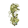
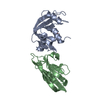
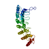
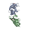
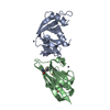
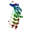
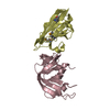
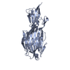
- PDB-4hbd: Crystal structure of KANK2 ankyrin repeats -
+
Open data
ID or keywords:
Loading...
-
Basic information
Entry
Database: PDB / ID: 4hbd
Title
Crystal structure of KANK2 ankyrin repeats
Components
KN motif and ankyrin repeat domain-containing protein 2
Keywords
PROTEIN BINDING / Structural Genomics Consortium / SGC
Function / homology
Function and homology information
negative regulation of vitamin D receptor signaling pathway / kidney epithelium development / podocyte cell migration / negative regulation of actin filament polymerization / negative regulation of intracellular estrogen receptor signaling pathway / regulation of Rho protein signal transduction / negative regulation of programmed cell death / negative regulation of G1/S transition of mitotic cell cycle / negative regulation of cell population proliferation / apoptotic process ...negative regulation of vitamin D receptor signaling pathway / kidney epithelium development / podocyte cell migration / negative regulation of actin filament polymerization / negative regulation of intracellular estrogen receptor signaling pathway / regulation of Rho protein signal transduction / negative regulation of programmed cell death / negative regulation of G1/S transition of mitotic cell cycle / negative regulation of cell population proliferation / apoptotic process / DNA-templated transcription / negative regulation of transcription by RNA polymerase II / mitochondrion / cytoplasm / cytosol Similarity search - Function
KNmotifandankyrinrepeatdomain-containingprotein2 / Ankyrin repeat domain-containing protein 25 / Matrix-remodeling-associated protein 3 / SRC-1- ...Ankyrin repeat domain-containing protein 25 / Matrix-remodeling-associated protein 3 / SRC-1-interacting protein / SRC1-interacting protein
Mass: 30162.223 Da / Num. of mol.: 1 / Fragment: UNP residues 578-832 Source method: isolated from a genetically manipulated source Source: (gene. exp.) Homo sapiens (human) / Gene: KANK2, ANKRD25, KIAA1518, MXRA3, SIP / Plasmid: pET28-MHL / Production host: Escherichia coli (E. coli) / Strain (production host): BL21-V2R-pRARE2 / References: UniProt: Q63ZY3
Resolution: 1.72→75.031 Å / Num. all: 31276 / Num. obs: 31276 / % possible obs: 100 % / Redundancy: 7.1 % / Rsym value: 0.072 / Net I/σ(I): 17.7
Reflection shell
Diffraction-ID: 1
Resolution (Å)
Redundancy (%)
Rmerge(I) obs
Mean I/σ(I) obs
Num. measured all
Num. unique all
Rsym value
% possible all
1.72-1.81
7.3
0.961
0.8
32689
4483
0.961
100
1.81-1.92
7.3
0.602
1.2
31103
4262
0.602
100
1.92-2.06
7.3
0.324
2.3
29168
4000
0.324
100
2.06-2.22
7.3
0.187
3.9
27039
3720
0.187
100
2.22-2.43
7.3
0.117
6.4
24975
3439
0.117
100
2.43-2.72
7.2
0.084
8.9
22675
3135
0.084
100
2.72-3.14
7.1
0.055
13.3
19954
2810
0.055
100
3.14-3.85
6.9
0.037
18
16420
2390
0.037
100
3.85-5.44
6.7
0.031
18.9
12699
1903
0.031
100
5.44-37.515
6
0.033
12.4
6829
1134
0.033
99.7
-
Phasing
Phasing
Method: molecular replacement
-
Processing
Software
Name
Version
Classification
NB
SCALA
3.3.20
datascaling
SHELX
phasing
REFMAC
5.7.0027
refinement
PDB_EXTRACT
3.11
dataextraction
XDS
datareduction
PHASER
phasing
Refinement
Method to determine structure: MOLECULAR REPLACEMENT Starting model: UNPUBLISHED MODEL OF SAME PROTEIN BUT DIFFERENT CRYSTAL DIMENSIONS (P21212; A,B,C=61.46,63.59,163.08 FOR DERIVATIVE (IODIDE?, DIFFRACTION INTENSITIES INCLUDED). THAT STRUCTURE WAS ...Starting model: UNPUBLISHED MODEL OF SAME PROTEIN BUT DIFFERENT CRYSTAL DIMENSIONS (P21212; A,B,C=61.46,63.59,163.08 FOR DERIVATIVE (IODIDE?, DIFFRACTION INTENSITIES INCLUDED). THAT STRUCTURE WAS SOLVED WITH SHELX, SIRAS (ISOMORPHOUS "NATIVE" DATA NOT PROVIDED AS SAD DOES ALSO WORK) Resolution: 1.72→37.54 Å / Cor.coef. Fo:Fc: 0.965 / Cor.coef. Fo:Fc free: 0.957 / WRfactor Rfree: 0.1973 / WRfactor Rwork: 0.1619 / Occupancy max: 1 / Occupancy min: 0.3 / FOM work R set: 0.8638 / SU B: 4.465 / SU ML: 0.073 / SU R Cruickshank DPI: 0.0974 / SU Rfree: 0.0967 / Cross valid method: THROUGHOUT / σ(F): 0 / ESU R: 0.097 / ESU R Free: 0.097 / Stereochemistry target values: MAXIMUM LIKELIHOOD Details: HYDROGENS HAVE BEEN ADDED IN THE RIDING POSITIONS. U VALUES: WITH TLS ADDED. ELECTRON DENSITY AT RESIDUES HIS-677 AND HIS-782 IS NOT CONSISTENT WITH THAT RESIDUE TYPE.
Rfactor
Num. reflection
% reflection
Selection details
Rfree
0.2072
1583
5.1 %
RANDOM
Rwork
0.1765
-
-
-
obs
0.1781
31215
99.98 %
-
Solvent computation
Ion probe radii: 0.8 Å / Shrinkage radii: 0.8 Å / VDW probe radii: 1.2 Å / Solvent model: MASK
In the structure databanks used in Yorodumi, some data are registered as the other names, "COVID-19 virus" and "2019-nCoV". Here are the details of the virus and the list of structure data.
Jan 31, 2019. EMDB accession codes are about to change! (news from PDBe EMDB page)
EMDB accession codes are about to change! (news from PDBe EMDB page)
The allocation of 4 digits for EMDB accession codes will soon come to an end. Whilst these codes will remain in use, new EMDB accession codes will include an additional digit and will expand incrementally as the available range of codes is exhausted. The current 4-digit format prefixed with “EMD-” (i.e. EMD-XXXX) will advance to a 5-digit format (i.e. EMD-XXXXX), and so on. It is currently estimated that the 4-digit codes will be depleted around Spring 2019, at which point the 5-digit format will come into force.
The EM Navigator/Yorodumi systems omit the EMD- prefix.
Related info.:Q: What is EMD? / ID/Accession-code notation in Yorodumi/EM Navigator
Yorodumi is a browser for structure data from EMDB, PDB, SASBDB, etc.
This page is also the successor to EM Navigator detail page, and also detail information page/front-end page for Omokage search.
The word "yorodu" (or yorozu) is an old Japanese word meaning "ten thousand". "mi" (miru) is to see.
Related info.:EMDB / PDB / SASBDB / Comparison of 3 databanks / Yorodumi Search / Aug 31, 2016. New EM Navigator & Yorodumi / Yorodumi Papers / Jmol/JSmol / Function and homology information / Changes in new EM Navigator and Yorodumi
 Movie
Movie Controller


 Open data
 Basic information
 Components
 Keywords
 Function and homology information
 Homo sapiens (human)
 X-RAY DIFFRACTION /
  Authors
 Citation
 Structure visualization
 Downloads & links
 Other downloads
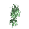

 PDBj
PDBj

 Assembly
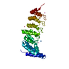


 Num. of mol.: 9 / Source method: obtained synthetically
 Mass: 18.015 Da / Num. of mol.: 168 / Source method: isolated from a natural source / Formula: H2O
 Sample preparation
 Processing