 Movie
Movie Controller


+ Open data

- Basic information
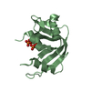
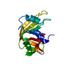
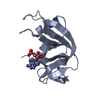
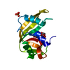
| Entry | Database: PDB / ID: 1kf2 | ||||||
|---|---|---|---|---|---|---|---|
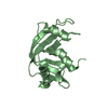
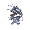
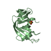
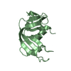
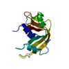
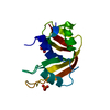
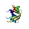
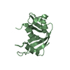
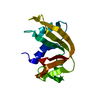
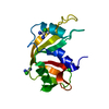
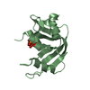
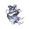
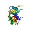
| Title | Atomic Resolution Structure of RNase A at pH 5.2 | ||||||
 Components Components | pancreatic ribonuclease | ||||||
 Keywords Keywords | HYDROLASE / RNase A / titration / pH / crystal / soaking | ||||||
| Function / homology |  Function and homology information Function and homology informationpancreatic ribonuclease / ribonuclease A activity / RNA nuclease activity / nucleic acid binding / defense response to Gram-positive bacterium / hydrolase activity / extracellular region Similarity search - Function | ||||||
| Biological species |  | ||||||
| Method |  X-RAY DIFFRACTION / SYNCHROTRON / MOLECULAR REPLACEMENT / Resolution: 1.1 Å X-RAY DIFFRACTION / SYNCHROTRON / MOLECULAR REPLACEMENT / Resolution: 1.1 Å | ||||||
 Authors Authors | Berisio, R. / Sica, F. / Lamzin, V.S. / Wilson, K.S. / Zagari, A. / Mazzarella, L. | ||||||
 Citation Citation | Journal: Acta Crystallogr.,Sect.D / Year: 2002 Title: Atomic resolution structures of ribonuclease A at six pH values. Authors: Berisio, R. / Sica, F. / Lamzin, V.S. / Wilson, K.S. / Zagari, A. / Mazzarella, L. #1: Journal: J.Mol.Biol. / Year: 1999Title: Protein titration in the crystal state Authors: Berisio, R. / Lamzin, V.S. / Sica, F. / Wilson, K.S. / Zagari, A. / Mazzarella, L. | ||||||
| History |
|
- Structure visualization
| Structure viewer | Molecule: MolmilJmol/JSmol |
|---|
- Downloads & links
-Download
| PDBx/mmCIF format | 1kf2.cif.gz | 75.6 KB | Display | PDBx/mmCIF format |
|---|---|---|---|---|
| PDB format | pdb1kf2.ent.gz | 56.5 KB | Display | PDB format |
| PDBx/mmJSON format | 1kf2.json.gz | Tree view | PDBx/mmJSON format | |
| Others |  Other downloads Other downloads |
-Validation report
| Arichive directory | https://data.pdbj.org/pub/pdb/validation_reports/kf/1kf2ftp://data.pdbj.org/pub/pdb/validation_reports/kf/1kf2 | HTTPS FTP |
|---|
-Related structure data
| Related structure data |  1kf3C 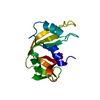 1kf4C 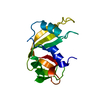 1kf5C 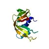 1kf7C 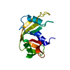 1kf8C  7rsaS C: citing same article ( S: Starting model for refinement |
|---|---|
| Similar structure data |


-Links
 PDBj
PDBj

- Assembly
| Deposited unit | 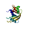
| ||||||||
|---|---|---|---|---|---|---|---|---|---|
| 1 |
| ||||||||
| Unit cell |
|
-Components
| #1: Protein | Mass: 13708.326 Da / Num. of mol.: 1 / Source method: isolated from a natural source / Source: (natural) |
|---|---|
| #2: Chemical | ChemComp-SO4 /   Mass: 96.063 Da / Num. of mol.: 1 / Source method: obtained synthetically / Formula: SO4 Mass: 96.063 Da / Num. of mol.: 1 / Source method: obtained synthetically / Formula: SO4 |
| #3: Water | ChemComp-HOH /  Mass: 18.015 Da / Num. of mol.: 244 / Source method: isolated from a natural source / Formula: H2O Mass: 18.015 Da / Num. of mol.: 244 / Source method: isolated from a natural source / Formula: H2O |
| Has protein modification | Y |
-Experimental details
-Experiment
| Experiment | Method: X-RAY DIFFRACTION / Number of used crystals: 1 |
|---|
- Sample preparation
| Crystal | Density Matthews: 2.19 Å3/Da / Density % sol: 43.74 % | ||||||||||||||||||||||||
|---|---|---|---|---|---|---|---|---|---|---|---|---|---|---|---|---|---|---|---|---|---|---|---|---|---|
| Crystal grow | Temperature: 298 K / Method: liquid diffusion / pH: 5.2 Details: 2-propanol, pH 5.2, LIQUID DIFFUSION, temperature 298K | ||||||||||||||||||||||||
| Crystal grow | *PLUS Temperature: 20 ℃ / Method: batch method / Details: Tilton Jr., R.F., (1992) Biochemistry, 31, 2469. | ||||||||||||||||||||||||
| Components of the solutions | *PLUS
|
-Data collection
| Diffraction | Mean temperature: 298 K |
|---|---|
| Diffraction source | Source: SYNCHROTRON / Site: EMBL/DESY, HAMBURG  / Beamline: X11 / Beamline: X11 |
| Detector | Type: MARRESEARCH / Detector: AREA DETECTOR |
| Radiation | Protocol: SINGLE WAVELENGTH / Monochromatic (M) / Laue (L): M / Scattering type: x-ray |
| Radiation wavelength | Relative weight: 1 |
| Reflection | Resolution: 1.1→30 Å / Num. all: 46617 / Num. obs: 46617 / % possible obs: 96.7 % / Observed criterion σ(I): 0 / Redundancy: 4.1 % / Biso Wilson estimate: 12 Å2 / Rmerge(I) obs: 0.034 / Net I/σ(I): 16 |
| Reflection shell | Resolution: 1.1→1.12 Å / Rmerge(I) obs: 0.329 / Mean I/σ(I) obs: 4.1 / % possible all: 89.4 |
| Reflection | *PLUS Highest resolution: 1.1 Å / Lowest resolution: 30 Å / Num. obs: 46613 |
- Processing
| Software |
| ||||||||||||
|---|---|---|---|---|---|---|---|---|---|---|---|---|---|
| Refinement | Method to determine structure: MOLECULAR REPLACEMENT Starting model: pdb entry 7rsa Resolution: 1.1→30 Å / σ(F): 0 / Stereochemistry target values: ENGH AND HUBER Details: PLEASE NOTE: ASUL 325, BSUL 325, AND CHOH 183 ARE ALTERNATE CONFORMATIONS. CHOH 173 IS ALTERNATIVE ONLY TO THE A CONFORMER OF HIS119. CHOH 150 IS ALTERNATIVE ONLY TO THE A CONFORMER OF ASN103.
| ||||||||||||
| Refinement step | Cycle: LAST / Resolution: 1.1→30 Å
| ||||||||||||
| Refine LS restraints |
| ||||||||||||
| LS refinement shell | Resolution: 1.1→1.14 Å /
| ||||||||||||
| Software | *PLUS Name: SHELXL-96 / Classification: refinement | ||||||||||||
| Refinement | *PLUS Lowest resolution: 30 Å / σ(F): 0 | ||||||||||||
| Solvent computation | *PLUS | ||||||||||||
| Displacement parameters | *PLUS | ||||||||||||
| Refine LS restraints | *PLUS
|
