 Movie
Movie Controller

[English] 日本語
 Yorodumi
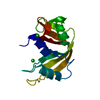
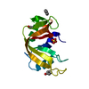
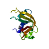
- PDB-4srn: STRUCTURAL CHANGES THAT ACCOMPANY THE REDUCED CATALYTIC EFFICIENC... -
+ Open data

- Basic information
| Entry | Database: PDB / ID: 4srn | ||||||
|---|---|---|---|---|---|---|---|
| Title | STRUCTURAL CHANGES THAT ACCOMPANY THE REDUCED CATALYTIC EFFICIENCY OF TWO SEMISYNTHETIC RIBONUCLEASE ANALOGS | ||||||
 Components Components | (RIBONUCLEASE A) x 2 | ||||||
 Keywords Keywords | HYDROLASE(NUCLEIC ACID / RNA) | ||||||
| Function / homology |  Function and homology information Function and homology informationpancreatic ribonuclease / ribonuclease A activity / RNA nuclease activity / nucleic acid binding / defense response to Gram-positive bacterium / hydrolase activity / extracellular region Similarity search - Function | ||||||
| Biological species |  | ||||||
| Method |  X-RAY DIFFRACTION / Resolution: 2 Å X-RAY DIFFRACTION / Resolution: 2 Å | ||||||
 Authors Authors | deMel, V.S.J. / Martin, P.D. / Doscher, M.S. / Edwards, B.F.P. | ||||||
 Citation Citation | Journal: J.Biol.Chem. / Year: 1992 Title: Structural changes that accompany the reduced catalytic efficiency of two semisynthetic ribonuclease analogs. Authors: deMel, V.S. / Martin, P.D. / Doscher, M.S. / Edwards, B.F. | ||||||
| History |
|
- Structure visualization
| Structure viewer | Molecule: MolmilJmol/JSmol |
|---|
- Downloads & links
-Download
| PDBx/mmCIF format | 4srn.cif.gz | 39 KB | Display | PDBx/mmCIF format |
|---|---|---|---|---|
| PDB format | pdb4srn.ent.gz | 26.9 KB | Display | PDB format |
| PDBx/mmJSON format | 4srn.json.gz | Tree view | PDBx/mmJSON format | |
| Others |  Other downloads Other downloads |
-Validation report
| Arichive directory | https://data.pdbj.org/pub/pdb/validation_reports/sr/4srnftp://data.pdbj.org/pub/pdb/validation_reports/sr/4srn | HTTPS FTP |
|---|
-Related structure data


-Links
 PDBj
PDBj

- Assembly
| Deposited unit | 
| ||||||||
|---|---|---|---|---|---|---|---|---|---|
| 1 |
| ||||||||
| Unit cell |
| ||||||||
| Atom site foot note | 1: CIS PROLINE - PRO A 93 |
-Components
| #1: Protein | Mass: 12494.965 Da / Num. of mol.: 1 Source method: isolated from a genetically manipulated source Source: (gene. exp.) |
|---|---|
| #2: Protein/peptide | Mass: 1187.365 Da / Num. of mol.: 1 Source method: isolated from a genetically manipulated source Source: (gene. exp.) |
| #3: Chemical | ChemComp-SO4 /   Mass: 96.063 Da / Num. of mol.: 1 / Source method: obtained synthetically / Formula: SO4 Mass: 96.063 Da / Num. of mol.: 1 / Source method: obtained synthetically / Formula: SO4 |
| #4: Water | ChemComp-HOH /  Mass: 18.015 Da / Num. of mol.: 111 / Source method: isolated from a natural source / Formula: H2O Mass: 18.015 Da / Num. of mol.: 111 / Source method: isolated from a natural source / Formula: H2O |
| Compound details | FEATURES OF THE STRUCTURE: 1. IN SPITE OF AN EIGHT-RESIDUE REDUNDANCY BETWEEN RNASE 1 - 118 AND ...FEATURES OF THE STRUCTURE: 1. IN SPITE OF AN EIGHT-RESIDUE REDUNDANCY |
| Has protein modification | Y |
| Nonpolymer details | A TOTAL OF 111 WATER MOLECULES WERE INCLUDED. A SULFATE ANION OCCURS IN THE ACTIVE SITE AND WAS ...A TOTAL OF 111 WATER MOLECULES WERE INCLUDED. A SULFATE ANION OCCURS IN THE ACTIVE SITE AND WAS ALSO REFINED AS PART OF THE STRUCTURE. |
-Experimental details
-Experiment
| Experiment | Method: X-RAY DIFFRACTION |
|---|
- Sample preparation
| Crystal | Density Matthews: 2.87 Å3/Da / Density % sol: 57.12 % | ||||||||||||||||||||||||||||||||||||||||||
|---|---|---|---|---|---|---|---|---|---|---|---|---|---|---|---|---|---|---|---|---|---|---|---|---|---|---|---|---|---|---|---|---|---|---|---|---|---|---|---|---|---|---|---|
| Crystal grow | Details: THE MOTHER LIQUOR WAS 80 PERCENT SATURATED AMMONIUM SULFATE, PH 5.2. | ||||||||||||||||||||||||||||||||||||||||||
| Crystal grow | *PLUS pH: 5.2 / Method: macro seeding | ||||||||||||||||||||||||||||||||||||||||||
| Components of the solutions | *PLUS
|
-Data collection
| Radiation | Scattering type: x-ray |
|---|---|
| Radiation wavelength | Relative weight: 1 |
| Reflection | Observed criterion σ(F): 2 |
| Reflection | *PLUS Highest resolution: 2 Å / Num. obs: 10236 / % possible obs: 90.7 % / Rmerge(I) obs: 0.051 |
- Processing
| Software |
| ||||||||||||||||||||||||||||||||||||||||||||||||||||||||||||
|---|---|---|---|---|---|---|---|---|---|---|---|---|---|---|---|---|---|---|---|---|---|---|---|---|---|---|---|---|---|---|---|---|---|---|---|---|---|---|---|---|---|---|---|---|---|---|---|---|---|---|---|---|---|---|---|---|---|---|---|---|---|
| Refinement | Resolution: 2→5 Å / Rfactor Rwork: 0.172 / Rfactor obs: 0.172 | ||||||||||||||||||||||||||||||||||||||||||||||||||||||||||||
| Refinement step | Cycle: LAST / Resolution: 2→5 Å
| ||||||||||||||||||||||||||||||||||||||||||||||||||||||||||||
| Refine LS restraints |
| ||||||||||||||||||||||||||||||||||||||||||||||||||||||||||||
| Software | *PLUS Name: X-PLOR/PROLSQ / Classification: refinement | ||||||||||||||||||||||||||||||||||||||||||||||||||||||||||||
| Refinement | *PLUS Num. reflection obs: 10236 / σ(F): 2 / Rfactor obs: 0.172 | ||||||||||||||||||||||||||||||||||||||||||||||||||||||||||||
| Solvent computation | *PLUS | ||||||||||||||||||||||||||||||||||||||||||||||||||||||||||||
| Displacement parameters | *PLUS | ||||||||||||||||||||||||||||||||||||||||||||||||||||||||||||
| Refine LS restraints | *PLUS
|
