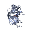
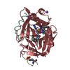
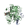
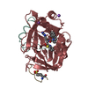
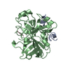
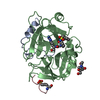
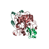
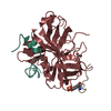
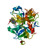
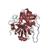
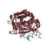
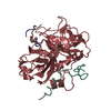
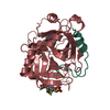
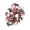
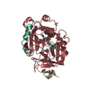
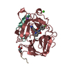
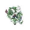
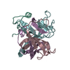
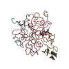
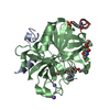
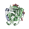
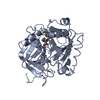
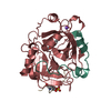
SHEET DETERMINATION METHOD: DSSP THE SHEETS PRESENTED AS "BB" IN EACH CHAIN ON SHEET RECORDS BELOW ... SHEET DETERMINATION METHOD: DSSP THE SHEETS PRESENTED AS "BB" IN EACH CHAIN ON SHEET RECORDS BELOW IS ACTUALLY AN 6-STRANDED BARREL THIS IS REPRESENTED BY A 7-STRANDED SHEET IN WHICH THE FIRST AND LAST STRANDS ARE IDENTICAL.
Mass: 18.015 Da / Num. of mol.: 388 / Source method: isolated from a natural source / Formula: H2O
-
Details
Compound details
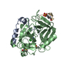
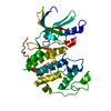
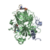
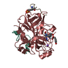
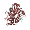
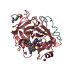
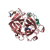
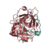
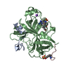
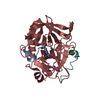
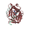
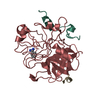
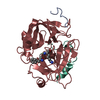
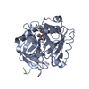
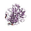
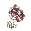
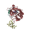
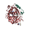
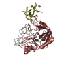
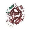
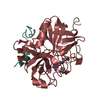
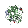
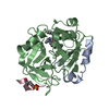
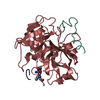
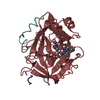
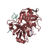
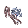
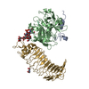
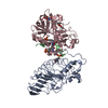
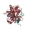
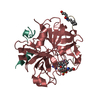
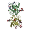
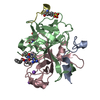
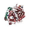
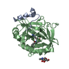
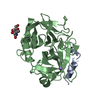
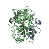
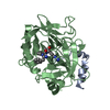
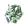
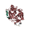
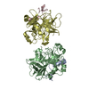
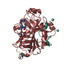
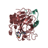
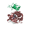
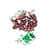
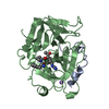
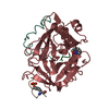
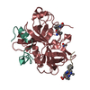
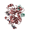
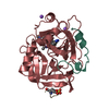
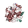
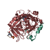
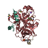
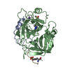
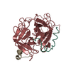
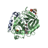
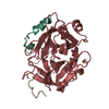
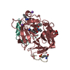
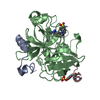
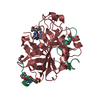
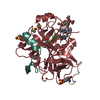
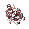
FUNCTION: THROMBIN, WHICH CLEAVES BONDS AFTER ARG AND LYS, CONVERTS FIBRINOGEN TO FIBRIN AND ...FUNCTION: THROMBIN, WHICH CLEAVES BONDS AFTER ARG AND LYS, CONVERTS FIBRINOGEN TO FIBRIN AND ACTIVATES FACTORS V, VII, VIII, XIII, AND, IN COMPLEX WITH THROMBOMODULIN, PROTEIN C. CATALYTIC ACTIVITY: PREFERENTIAL CLEAVAGE: ARG-|-GLY; ACTIVATES FIBRINOGEN TO FIBRIN AND RELEASES FIBRINOPEPTIDE A AND B. SUBCELLULAR LOCATION: EXTRACELLULAR. TISSUE SPECIFICITY: EXPRESSED BY THE LIVER AND SECRETED IN PLASMA. DISEASE: DEFECTS IN F2 ARE THE CAUSE OF VARIOUS FORMS OF DYSPROTHROMBINEMIA [MIM:176930]. SIMILARITY: BELONGS TO THE PEPTIDASE S1 FAMILY. SIMILARITY: CONTAINS 1 GAMMA-CARBOXY-GLUTAMATE DOMAIN (GLA) DOMAIN. SIMILARITY: CONTAINS 2 KRINGLE DOMAINS. FUNCTION: HIRUDIN IS A POTENT THROMBIN-SPECIFIC PROTEASE INHIBITOR. IT FORMS A STABLE NON-COVALENT COMPLEX WITH ALPHA- THROMBIN, THEREBY ABOLISHING ITS ABILITY TO CLEAVE FIBRINOGEN. SUBCELLULAR LOCATION: SECRETED.
-
Experimental details
-
Experiment
Experiment
Method: X-RAY DIFFRACTION / Number of used crystals: 1
-
Sample preparation
Crystal
Density Matthews: 2.5 Å3/Da / Density % sol: 50.75 %
Resolution: 2.2→27.21 Å / Cor.coef. Fo:Fc: 0.957 / Cor.coef. Fo:Fc free: 0.895 / SU B: 5.98 / SU ML: 0.154 / Cross valid method: THROUGHOUT / ESU R: 0.272 / ESU R Free: 0.247 / Stereochemistry target values: MAXIMUM LIKELIHOOD Details: HYDROGENS HAVE BEEN ADDED IN THE RIDING POSITIONS. DISORDER AT TWO-FOLD; ARG B 75 CLASHES. THE OCCUPANCY OF SIDE CHAIN ATOMS WAS SET TO 0 FOR REFINEMENT. THESE ATOMS REMOVED FOR DURING PDB ANNOTATION.
Rfactor
Num. reflection
% reflection
Selection details
Rfree
0.265
876
5.1 %
RANDOM
Rwork
0.166
-
-
-
obs
0.171
16409
97.6 %
-
Solvent computation
Ion probe radii: 0.8 Å / Shrinkage radii: 0.8 Å / VDW probe radii: 1.2 Å / Solvent model: BABINET MODEL WITH MASK
 Movie
Movie Controller


 Open data
 Basic information
 Components
 Keywords
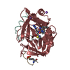
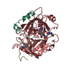
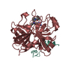
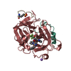
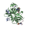
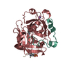
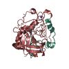
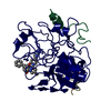
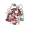
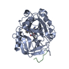
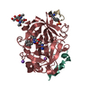
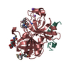
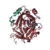
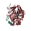
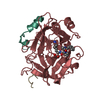
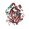
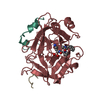
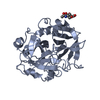
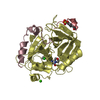
 SERINE PROTEASE /
  Function and homology information

 Authors
 Citation
 Structure visualization
 Downloads & links
 Other downloads


 PDBj
PDBj


 Assembly
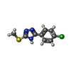


 Mass: 225.698 Da / Num. of mol.: 1 / Source method: obtained synthetically / Formula: C9H8ClN3S
 Mass: 78.133 Da / Num. of mol.: 3 / Source method: obtained synthetically / Formula: C2H6OS / Comment: DMSO, precipitant*YM
 Sample preparation
 Processing