 Movie
Movie Controller

[English] 日本語
 Yorodumi
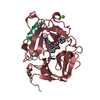
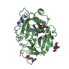
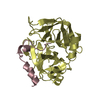
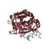
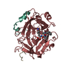
- PDB-1dm4: SER195ALA MUTANT OF HUMAN THROMBIN COMPLEXED WITH FIBRINOPEPTIDE ... -
+ Open data

- Basic information
| Entry | Database: PDB / ID: 1dm4 | ||||||
|---|---|---|---|---|---|---|---|
| Title | SER195ALA MUTANT OF HUMAN THROMBIN COMPLEXED WITH FIBRINOPEPTIDE A (7-16) | ||||||
 Components Components |
| ||||||
 Keywords Keywords | HYDROLASE / MUTANT THROMBIN / RESIDUAL CATALYTIC ACTIVITY / FIBRINOPEPTIDE A | ||||||
| Function / homology |  Function and homology information Function and homology informationblood coagulation, common pathway / induction of bacterial agglutination / fibrinogen complex / platelet alpha granule / Regulation of TLR by endogenous ligand / MyD88 deficiency (TLR2/4) / positive regulation of heterotypic cell-cell adhesion / IRAK4 deficiency (TLR2/4) / MyD88:MAL(TIRAP) cascade initiated on plasma membrane / plasminogen activation ...blood coagulation, common pathway / induction of bacterial agglutination / fibrinogen complex / platelet alpha granule / Regulation of TLR by endogenous ligand / MyD88 deficiency (TLR2/4) / positive regulation of heterotypic cell-cell adhesion / IRAK4 deficiency (TLR2/4) / MyD88:MAL(TIRAP) cascade initiated on plasma membrane / plasminogen activation / extracellular matrix structural constituent / : / thrombospondin receptor activity / p130Cas linkage to MAPK signaling for integrins / thrombin / thrombin-activated receptor signaling pathway / Defective factor XII causes hereditary angioedema / positive regulation of peptide hormone secretion / negative regulation of astrocyte differentiation / regulation of blood coagulation / neutrophil-mediated killing of gram-negative bacterium / positive regulation of phospholipase C-activating G protein-coupled receptor signaling pathway / Defective F8 cleavage by thrombin / ligand-gated ion channel signaling pathway / Platelet Aggregation (Plug Formation) / GRB2:SOS provides linkage to MAPK signaling for Integrins / positive regulation of collagen biosynthetic process / positive regulation of exocytosis / negative regulation of platelet activation / negative regulation of blood coagulation / negative regulation of fibrinolysis / blood coagulation, fibrin clot formation / positive regulation of blood coagulation / protein polymerization / negative regulation of endothelial cell apoptotic process / positive regulation of vasoconstriction / Integrin cell surface interactions / Transport of gamma-carboxylated protein precursors from the endoplasmic reticulum to the Golgi apparatus / : / Gamma-carboxylation of protein precursors / Removal of aminoterminal propeptides from gamma-carboxylated proteins / negative regulation of extrinsic apoptotic signaling pathway via death domain receptors / regulation of cytosolic calcium ion concentration / fibrinolysis / : / negative regulation of proteolysis / Integrin signaling / positive regulation of substrate adhesion-dependent cell spreading / negative regulation of cytokine production involved in inflammatory response / platelet alpha granule lumen / Regulation of Complement cascade / positive regulation of release of sequestered calcium ion into cytosol / acute-phase response / cell-matrix adhesion / Cell surface interactions at the vascular wall / Peptide ligand-binding receptors / positive regulation of protein secretion / growth factor activity / positive regulation of receptor signaling pathway via JAK-STAT / Post-translational protein phosphorylation / lipopolysaccharide binding / Signaling by high-kinase activity BRAF mutants / platelet activation / MAP2K and MAPK activation / positive regulation of protein localization to nucleus / response to calcium ion / response to wounding / Golgi lumen / platelet aggregation / Regulation of Insulin-like Growth Factor (IGF) transport and uptake by Insulin-like Growth Factor Binding Proteins (IGFBPs) / positive regulation of reactive oxygen species metabolic process / blood coagulation / positive regulation of insulin secretion / Signaling by RAF1 mutants / Signaling by moderate kinase activity BRAF mutants / Paradoxical activation of RAF signaling by kinase inactive BRAF / Signaling downstream of RAS mutants / Signaling by BRAF and RAF1 fusions / Platelet degranulation / regulation of cell shape / antimicrobial humoral immune response mediated by antimicrobial peptide / heparin binding / extracellular vesicle / extracellular matrix / Thrombin signalling through proteinase activated receptors (PARs) / positive regulation of cell growth / ER-Phagosome pathway / protein-containing complex assembly / blood microparticle / G alpha (q) signalling events / adaptive immune response / positive regulation of ERK1 and ERK2 cascade / protein-macromolecule adaptor activity / positive regulation of phosphatidylinositol 3-kinase/protein kinase B signal transduction / cell surface receptor signaling pathway / endoplasmic reticulum lumen / Amyloid fiber formation / receptor ligand activity / serine-type endopeptidase activity / external side of plasma membrane Similarity search - Function | ||||||
| Biological species |  Homo sapiens (human) Homo sapiens (human) | ||||||
| Method |  X-RAY DIFFRACTION / Resolution: 2.5 Å X-RAY DIFFRACTION / Resolution: 2.5 Å | ||||||
 Authors Authors | Krishnan, R. / Sadler, E.J. / Tulinsky, A. | ||||||
 Citation Citation | Journal: Acta Crystallogr.,Sect.D / Year: 2000 Title: Structure of the Ser195Ala mutant of human alpha--thrombin complexed with fibrinopeptide A(7--16): evidence for residual catalytic activity. Authors: Krishnan, R. / Sadler, J.E. / Tulinsky, A. | ||||||
| History |
|
- Structure visualization
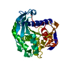
| Structure viewer | Molecule: MolmilJmol/JSmol |
|---|
- Downloads & links
-Download
| PDBx/mmCIF format | 1dm4.cif.gz | 75.6 KB | Display | PDBx/mmCIF format |
|---|---|---|---|---|
| PDB format | pdb1dm4.ent.gz | 56.5 KB | Display | PDB format |
| PDBx/mmJSON format | 1dm4.json.gz | Tree view | PDBx/mmJSON format | |
| Others |  Other downloads Other downloads |
-Validation report
| Arichive directory | https://data.pdbj.org/pub/pdb/validation_reports/dm/1dm4ftp://data.pdbj.org/pub/pdb/validation_reports/dm/1dm4 | HTTPS FTP |
|---|
-Related structure data
| Similar structure data |
|---|


-Links
 PDBj
PDBj


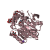
- Assembly
| Deposited unit | 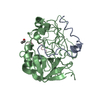
| ||||||||
|---|---|---|---|---|---|---|---|---|---|
| 1 |
| ||||||||
| 2 | 
| ||||||||
| Unit cell |
|
-Components
| #1: Protein/peptide | Mass: 3939.340 Da / Num. of mol.: 1 Source method: isolated from a genetically manipulated source Source: (gene. exp.) Homo sapiens (human) / References: UniProt: P00734 |
|---|---|
| #2: Protein | Mass: 29921.414 Da / Num. of mol.: 1 / Mutation: S195A Source method: isolated from a genetically manipulated source Source: (gene. exp.) Homo sapiens (human) / References: UniProt: P00734 |
| #3: Protein/peptide | Mass: 1047.144 Da / Num. of mol.: 1 / Fragment: FPA / Source method: obtained synthetically / Details: CHEMICALLY SYNTHESIZED / References: UniProt: P02671*PLUS |
| #4: Water | ChemComp-HOH /  Mass: 18.015 Da / Num. of mol.: 149 / Source method: isolated from a natural source / Formula: H2O Mass: 18.015 Da / Num. of mol.: 149 / Source method: isolated from a natural source / Formula: H2O |
| Has protein modification | Y |
-Experimental details
-Experiment
| Experiment | Method: X-RAY DIFFRACTION / Number of used crystals: 1 |
|---|
- Sample preparation
| Crystal | Density Matthews: 2.94 Å3/Da / Density % sol: 58.19 % | ||||||||||||||||||||
|---|---|---|---|---|---|---|---|---|---|---|---|---|---|---|---|---|---|---|---|---|---|
| Crystal grow | pH: 7.5 Details: 1.4M SODIUM CITRATE, 1-2% ISOPROPANOL, 0.1M HEPES BUFFER, pH 7.50 | ||||||||||||||||||||
| Crystal grow | *PLUS Temperature: 277 K / pH: 7.5 / Method: vapor diffusion, hanging drop | ||||||||||||||||||||
| Components of the solutions | *PLUS
|
-Data collection
| Diffraction | Mean temperature: 123 K |
|---|---|
| Diffraction source | Source: ROTATING ANODE / Type: RIGAKU RU200 / Wavelength: 1.5418 |
| Detector | Type: RIGAKU RAXIS II / Detector: IMAGE PLATE / Date: Mar 7, 1996 |
| Radiation | Protocol: SINGLE WAVELENGTH / Monochromatic (M) / Laue (L): M / Scattering type: x-ray |
| Radiation wavelength | Wavelength: 1.5418 Å / Relative weight: 1 |
| Reflection | Resolution: 2.5→15 Å / Num. obs: 9561 / % possible obs: 71 % / Observed criterion σ(I): 2 / Redundancy: 2.28 % / Biso Wilson estimate: 25 Å2 / Rmerge(I) obs: 0.093 / Net I/σ(I): 15 |
| Reflection shell | Resolution: 2.5→2.75 Å / Redundancy: 1.8 % / Rmerge(I) obs: 0.19 / % possible all: 94.6 |
| Reflection | *PLUS Highest resolution: 2.5 Å / Lowest resolution: 15 Å / Observed criterion σ(I): 2 / Num. measured all: 21833 |
| Reflection shell | *PLUS % possible obs: 61 % |
- Processing
| Software |
| |||||||||||||||||||||||||||||||||||||||||||||||||||||||||||||||
|---|---|---|---|---|---|---|---|---|---|---|---|---|---|---|---|---|---|---|---|---|---|---|---|---|---|---|---|---|---|---|---|---|---|---|---|---|---|---|---|---|---|---|---|---|---|---|---|---|---|---|---|---|---|---|---|---|---|---|---|---|---|---|---|---|
| Refinement | Resolution: 2.5→7 Å / σ(F): 4 / Details: USED WEIGHTED FULL MATRIX LEAST SQUARES PROCEDURE. /
| |||||||||||||||||||||||||||||||||||||||||||||||||||||||||||||||
| Refinement step | Cycle: LAST / Resolution: 2.5→7 Å
| |||||||||||||||||||||||||||||||||||||||||||||||||||||||||||||||
| Refine LS restraints |
| |||||||||||||||||||||||||||||||||||||||||||||||||||||||||||||||
| Software | *PLUS Name: PROFFT / Classification: refinement | |||||||||||||||||||||||||||||||||||||||||||||||||||||||||||||||
| Refinement | *PLUS Rfactor Rwork: 0.169 | |||||||||||||||||||||||||||||||||||||||||||||||||||||||||||||||
| Solvent computation | *PLUS | |||||||||||||||||||||||||||||||||||||||||||||||||||||||||||||||
| Displacement parameters | *PLUS | |||||||||||||||||||||||||||||||||||||||||||||||||||||||||||||||
| Refine LS restraints | *PLUS
|
