 Movie
Movie Controller

[English] 日本語
 Yorodumi
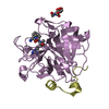
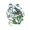
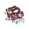
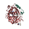
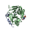
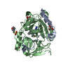
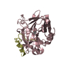
- PDB-1oyt: COMPLEX OF RECOMBINANT HUMAN THROMBIN WITH A DESIGNED FLUORINATED... -
+ Open data

- Basic information
| Entry | Database: PDB / ID: 1oyt | ||||||
|---|---|---|---|---|---|---|---|
| Title | COMPLEX OF RECOMBINANT HUMAN THROMBIN WITH A DESIGNED FLUORINATED INHIBITOR | ||||||
 Components Components |
| ||||||
 Keywords Keywords | HYDROLASE/HYDROLASE INHIBITOR / SERINE PROTEINASE / HYDROLASE-HYDROLASE INHIBITOR COMPLEX | ||||||
| Function / homology |  Function and homology information Function and homology information: / thrombospondin receptor activity / thrombin / thrombin-activated receptor signaling pathway / Defective factor XII causes hereditary angioedema / negative regulation of astrocyte differentiation / regulation of blood coagulation / neutrophil-mediated killing of gram-negative bacterium / positive regulation of phospholipase C-activating G protein-coupled receptor signaling pathway / Defective F8 cleavage by thrombin ...: / thrombospondin receptor activity / thrombin / thrombin-activated receptor signaling pathway / Defective factor XII causes hereditary angioedema / negative regulation of astrocyte differentiation / regulation of blood coagulation / neutrophil-mediated killing of gram-negative bacterium / positive regulation of phospholipase C-activating G protein-coupled receptor signaling pathway / Defective F8 cleavage by thrombin / ligand-gated ion channel signaling pathway / Platelet Aggregation (Plug Formation) / positive regulation of collagen biosynthetic process / negative regulation of platelet activation / negative regulation of blood coagulation / negative regulation of fibrinolysis / blood coagulation, fibrin clot formation / positive regulation of blood coagulation / Transport of gamma-carboxylated protein precursors from the endoplasmic reticulum to the Golgi apparatus / : / Gamma-carboxylation of protein precursors / Removal of aminoterminal propeptides from gamma-carboxylated proteins / regulation of cytosolic calcium ion concentration / fibrinolysis / : / negative regulation of proteolysis / negative regulation of cytokine production involved in inflammatory response / Regulation of Complement cascade / positive regulation of release of sequestered calcium ion into cytosol / acute-phase response / Cell surface interactions at the vascular wall / Peptide ligand-binding receptors / growth factor activity / positive regulation of receptor signaling pathway via JAK-STAT / serine-type endopeptidase inhibitor activity / lipopolysaccharide binding / platelet activation / positive regulation of protein localization to nucleus / response to wounding / Golgi lumen / Regulation of Insulin-like Growth Factor (IGF) transport and uptake by Insulin-like Growth Factor Binding Proteins (IGFBPs) / positive regulation of reactive oxygen species metabolic process / blood coagulation / positive regulation of insulin secretion / regulation of cell shape / antimicrobial humoral immune response mediated by antimicrobial peptide / heparin binding / Thrombin signalling through proteinase activated receptors (PARs) / positive regulation of cell growth / blood microparticle / G alpha (q) signalling events / positive regulation of phosphatidylinositol 3-kinase/protein kinase B signal transduction / cell surface receptor signaling pathway / endoplasmic reticulum lumen / receptor ligand activity / serine-type endopeptidase activity / signaling receptor binding / calcium ion binding / positive regulation of cell population proliferation / proteolysis / : / extracellular exosome / extracellular region / plasma membrane Similarity search - Function | ||||||
| Biological species |  Homo sapiens (human) Homo sapiens (human) Hirudo medicinalis (medicinal leech) Hirudo medicinalis (medicinal leech) | ||||||
| Method |  X-RAY DIFFRACTION / FOURIER SYNTHESIS / Resolution: 1.67 Å X-RAY DIFFRACTION / FOURIER SYNTHESIS / Resolution: 1.67 Å | ||||||
 Authors Authors | Banner, D.W. / Olsen, J.A. | ||||||
 Citation Citation | Journal: Angew.Chem.Int.Ed.Engl. / Year: 2003 Title: A Fluorine Scan of Thrombin Inhibitors to Map the Fluorophilicity/Fluorophobicity of an Enzyme Active Site: Evidence for CF...C=O Interactions. Authors: Olsen, J.A. / Banner, D.W. / Seiler, P. / Obst-Sander, U. / D'Arcy, A. / Stihle, M. / Mueller, K. / Diederich, F. #1: Journal: Protein Expr.Purif. / Year: 1997Title: Stable expression and purification of a secreted human recombinant prethrombin-2 and its activation to thrombin Authors: Russo, G. / Gast, A. / Schlaeger, E.J. / Angiolillo, A. / Pietropaolo, C. | ||||||
| History |
|
- Structure visualization
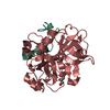
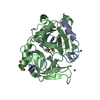
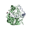
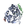
| Structure viewer | Molecule: MolmilJmol/JSmol |
|---|
- Downloads & links
-Download
| PDBx/mmCIF format | 1oyt.cif.gz | 85.7 KB | Display | PDBx/mmCIF format |
|---|---|---|---|---|
| PDB format | pdb1oyt.ent.gz | 62 KB | Display | PDB format |
| PDBx/mmJSON format | 1oyt.json.gz | Tree view | PDBx/mmJSON format | |
| Others |  Other downloads Other downloads |
-Validation report
| Arichive directory | https://data.pdbj.org/pub/pdb/validation_reports/oy/1oytftp://data.pdbj.org/pub/pdb/validation_reports/oy/1oyt | HTTPS FTP |
|---|
-Related structure data
| Similar structure data |
|---|


-Links
 PDBj
PDBj


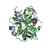
- Assembly
| Deposited unit | 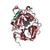
| |||||||||
|---|---|---|---|---|---|---|---|---|---|---|
| 1 |
| |||||||||
| Unit cell |
| |||||||||
| Components on special symmetry positions |
|
-Components
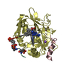
-Protein/peptide , 2 types, 2 molecules LI
| #1: Protein/peptide | Mass: 4096.534 Da / Num. of mol.: 1 Source method: isolated from a genetically manipulated source Source: (gene. exp.) Homo sapiens (human) / Gene: F2 / Cell (production host): OVARY / Production host:   Cricetulus griseus (Chinese hamster) / References: UniProt: P00734, thrombin Cricetulus griseus (Chinese hamster) / References: UniProt: P00734, thrombin |
|---|---|
| #3: Protein/peptide | 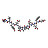  Type: Oligopeptide / Class: Anticoagulant, Antithrombotic / Mass: 1396.450 Da / Num. of mol.: 1 / Source method: obtained synthetically Type: Oligopeptide / Class: Anticoagulant, Antithrombotic / Mass: 1396.450 Da / Num. of mol.: 1 / Source method: obtained syntheticallyDetails: The sequence was chemically synthesized. The sequence is naturally found in Hirudo medicinalis (Medicinal leech). Source: (synth.) Hirudo medicinalis (medicinal leech)References: UniProt: P28506, N-terminal Asp des-amino Hirudin-3A |
-Protein , 1 types, 1 molecules H
| #2: Protein | Mass: 29780.219 Da / Num. of mol.: 1 Source method: isolated from a genetically manipulated source Source: (gene. exp.) Homo sapiens (human) / Gene: F2 / Cell (production host): OVARY / Production host: Cricetulus griseus (Chinese hamster) / References: UniProt: P00734, thrombin |
|---|
-Non-polymers , 4 types, 407 molecules 


| #4: Chemical | ChemComp-NA /  Mass: 22.990 Da / Num. of mol.: 1 / Source method: obtained synthetically / Formula: Na Mass: 22.990 Da / Num. of mol.: 1 / Source method: obtained synthetically / Formula: Na |
|---|---|
| #5: Chemical | ChemComp-CA /  Mass: 40.078 Da / Num. of mol.: 1 / Source method: obtained synthetically / Formula: Ca Mass: 40.078 Da / Num. of mol.: 1 / Source method: obtained synthetically / Formula: Ca |
| #6: Chemical | ChemComp-FSN / ( Mass: 407.461 Da / Num. of mol.: 1 / Source method: obtained synthetically / Formula: C23H24FN4O2 Mass: 407.461 Da / Num. of mol.: 1 / Source method: obtained synthetically / Formula: C23H24FN4O2 |
| #7: Water | ChemComp-HOH / Mass: 18.015 Da / Num. of mol.: 404 / Source method: isolated from a natural source / Formula: H2O |
-Experimental details
-Experiment
| Experiment | Method: X-RAY DIFFRACTION / Number of used crystals: 1 |
|---|
- Sample preparation
| Crystal | Density Matthews: 2.56 Å3/Da / Density % sol: 51.99 % | ||||||||||||||||||||||||||||
|---|---|---|---|---|---|---|---|---|---|---|---|---|---|---|---|---|---|---|---|---|---|---|---|---|---|---|---|---|---|
| Crystal grow | *PLUS pH: 7.4 / Method: unknown | ||||||||||||||||||||||||||||
| Components of the solutions | *PLUS
|
-Data collection
| Diffraction | Mean temperature: 100 K |
|---|---|
| Diffraction source | Source: ROTATING ANODE / Type: ENRAF-NONIUS FR591 / Wavelength: 1.5418 Å |
| Detector | Type: MAR scanner 345 mm plate / Detector: IMAGE PLATE / Date: May 24, 2002 / Details: Osmic mirrors |
| Radiation | Monochromator: OSMICS / Protocol: SINGLE WAVELENGTH / Monochromatic (M) / Laue (L): M / Scattering type: x-ray |
| Radiation wavelength | Wavelength: 1.5418 Å / Relative weight: 1 |
| Reflection | Resolution: 1.64→20 Å / Num. all: 44106 / Num. obs: 38758 / % possible obs: 87.9 % / Observed criterion σ(I): -3 / Redundancy: 7.33 % / Biso Wilson estimate: 21.8 Å2 / Rmerge(I) obs: 0.036 / Rsym value: 0.042 / Net I/σ(I): 33.43 |
| Reflection shell | Resolution: 1.64→1.74 Å / Redundancy: 6.25 % / Rmerge(I) obs: 0.16 / Mean I/σ(I) obs: 9.71 / Num. unique all: 7048 / Rsym value: 0.17 / % possible all: 53 |
| Reflection | *PLUS Highest resolution: 1.67 Å / Num. measured all: 284109 / Rmerge(I) obs: 0.033 |
| Reflection shell | *PLUS Rmerge(I) obs: 0.149 / Mean I/σ(I) obs: 9.7 |
- Processing
| Software |
| ||||||||||||||||||||||||||||||||||||
|---|---|---|---|---|---|---|---|---|---|---|---|---|---|---|---|---|---|---|---|---|---|---|---|---|---|---|---|---|---|---|---|---|---|---|---|---|---|
| Refinement | Method to determine structure: FOURIER SYNTHESIS Starting model: IN HOUSE THROMBIN STRUCTURES Resolution: 1.67→19.3 Å / Rfactor Rfree error: 0.005 / Isotropic thermal model: RESTRAINED / Cross valid method: THROUGHOUT / σ(F): 0 / Stereochemistry target values: Engh & Huber Details: CHYMOTRYPSIN NUMBERING (RATHER THAN SEQUENTIAL) SYSTEM IS USED, BASED ON THE TOPOLOGICAL ALIGNMENT WITH THE STRUCTURE OF CHYMOTRYPSIN (W.BODE ET AL., 1989, EMBO J. 8, 3467-3475). Double ...Details: CHYMOTRYPSIN NUMBERING (RATHER THAN SEQUENTIAL) SYSTEM IS USED, BASED ON THE TOPOLOGICAL ALIGNMENT WITH THE STRUCTURE OF CHYMOTRYPSIN (W.BODE ET AL., 1989, EMBO J. 8, 3467-3475). Double confromations given as single conformer are: H chain Leu41, Arg50, Leu64, Val66, Leu85, Met106, Ser129B Leu130, Asp186. Gly186C has a second trace with psi of 186D + 180degrees. Missing residues H chain: 148 loop WTANVGK, residues Thr147 and Gln151 weak. Missing residues L chain: N-terminus TFGSGE, C-terminus DGR. Missing from 'hirugen' I chain: C-terminus EEYLQ. Hirugen corresponds to hirudin 55-65, is synthesized chemically, is not sulphated. Asp 55 has no amino group and thus is given as a succinic acid residue. Arg75 has 2 conformations. COORDINATES ARE GIVEN UP TO CB. WATERS 107 AND 35 INDICATE THE GUANIDINIUM POSITION ON THE 2-FOLD AXIS. Residues with some or all atoms at 0.5 occupancy are L chain: Asp1A, Lys14A, Arg14D, Ile14K, H chain: Leu41, Lys81, Lys110, Pro111, Arg126, Glu127, Ser129B, Thr147, Gln151, Asp186A, Lys186D, Glu192, Asn205, Lys236 Gln239, Lys240, Gln244, I chain Glu4, Pro6. Atoms with zero occupancy H chain Ser36A Og and Asn62 OD1. The sugar on Asn62 is not modelled (water molecules). All inhibitor atoms refine to B-values < 25. The weakest is O18. Between O18 and the NZ of K60F is a smear of density, not well fitted by water 233.
| ||||||||||||||||||||||||||||||||||||
| Solvent computation | Solvent model: FLAT MODEL / Bsol: 48.7734 Å2 / ksol: 0.312145 e/Å3 | ||||||||||||||||||||||||||||||||||||
| Displacement parameters | Biso mean: 17.5 Å2
| ||||||||||||||||||||||||||||||||||||
| Refine analyze | Luzzati coordinate error free: 0.2 Å / Luzzati sigma a free: 0.12 Å | ||||||||||||||||||||||||||||||||||||
| Refinement step | Cycle: LAST / Resolution: 1.67→19.3 Å
| ||||||||||||||||||||||||||||||||||||
| Refine LS restraints |
| ||||||||||||||||||||||||||||||||||||
| LS refinement shell | Resolution: 1.67→1.73 Å / Rfactor Rfree error: 0.019 / Total num. of bins used: 10
| ||||||||||||||||||||||||||||||||||||
| Xplor file |
| ||||||||||||||||||||||||||||||||||||
| Refine LS restraints | *PLUS
|
