Mass: 18.015 Da / Num. of mol.: 22 / Source method: isolated from a natural source / Formula: H2O
-
Details
Has protein modification
Y
-
Experimental details
-
Experiment
Experiment
Method: X-RAY DIFFRACTION / Number of used crystals: 1
-
Sample preparation
Crystal
Density Matthews: 2.52 Å3/Da / Density % sol: 51.22 %
Crystal grow
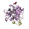
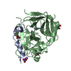
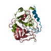
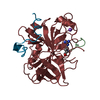
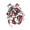
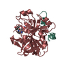
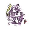
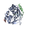
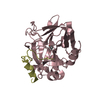
Temperature: 294 K / Method: batch mode Details: For complex formation, 2 mg human alpha-thrombin (7.1 mg/mL in 50% v/v glycerol) were mixed with 0.5 mM Compound 1 in DMSO and dialyzed overnight into a solution of 20 mM sodium citrate and ...Details: For complex formation, 2 mg human alpha-thrombin (7.1 mg/mL in 50% v/v glycerol) were mixed with 0.5 mM Compound 1 in DMSO and dialyzed overnight into a solution of 20 mM sodium citrate and 1.3 mM Na2SO4 (pH 5.8). After dialysis, another 0.5 mM compound were added and left to incubate at room temperature for 1 h. The protein was centrifuged for 5 min at 4 deg C and 8000x RCF. The pellet, including about 120 uL of buffer, was mixed with 100 uL of a solution containing 50 mM sodium phosphate (pH 7.3), 190 mM sodium chloride, and 0.2 mM Compound 1. The crystal used for data collection was grown from the JCSG+ screen in well A10. The crystal was grown at 20 deg C using a MRC 3-well plate and in a 150 + 150 nL drop with a reservoir of 0.2 M potassium formate and 20% w/v PEG-3350.
In the structure databanks used in Yorodumi, some data are registered as the other names, "COVID-19 virus" and "2019-nCoV". Here are the details of the virus and the list of structure data.
Jan 31, 2019. EMDB accession codes are about to change! (news from PDBe EMDB page)
EMDB accession codes are about to change! (news from PDBe EMDB page)
The allocation of 4 digits for EMDB accession codes will soon come to an end. Whilst these codes will remain in use, new EMDB accession codes will include an additional digit and will expand incrementally as the available range of codes is exhausted. The current 4-digit format prefixed with “EMD-” (i.e. EMD-XXXX) will advance to a 5-digit format (i.e. EMD-XXXXX), and so on. It is currently estimated that the 4-digit codes will be depleted around Spring 2019, at which point the 5-digit format will come into force.
The EM Navigator/Yorodumi systems omit the EMD- prefix.
Related info.:Q: What is EMD? / ID/Accession-code notation in Yorodumi/EM Navigator
Yorodumi is a browser for structure data from EMDB, PDB, SASBDB, etc.
This page is also the successor to EM Navigator detail page, and also detail information page/front-end page for Omokage search.
The word "yorodu" (or yorozu) is an old Japanese word meaning "ten thousand". "mi" (miru) is to see.
Related info.:EMDB / PDB / SASBDB / Comparison of 3 databanks / Yorodumi Search / Aug 31, 2016. New EM Navigator & Yorodumi / Yorodumi Papers / Jmol/JSmol / Function and homology information / Changes in new EM Navigator and Yorodumi
 Movie
Movie Controller


 Open data
 Basic information
 Components
 Keywords
 Function and homology information
 Homo sapiens (human)
 X-RAY DIFFRACTION /
  Authors
 Citation
 Structure visualization
 Downloads & links
 Other downloads
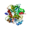


 PDBj
PDBj


 Assembly

 Type: D-saccharide, beta linking / Mass: 221.208 Da / Num. of mol.: 1


 Mass: 152.147 Da / Num. of mol.: 2 / Source method: obtained synthetically / Formula: C8H8O3
 Mass: 22.990 Da / Num. of mol.: 2 / Source method: obtained synthetically / Formula: Na
 Sample preparation
 / Beamline: I03 / Wavelength: 0.97625 Å
 Processing