 Movie
Movie Controller

[English] 日本語
 Yorodumi
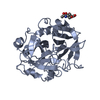
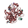
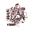
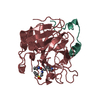
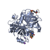
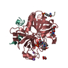
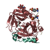
- PDB-1ycp: THE CRYSTAL STRUCTURE OF FIBRINOGEN-AA PEPTIDE 1-23 (F8Y) BOUND T... -
+ Open data

- Basic information
| Entry | Database: PDB / ID: 1ycp | ||||||
|---|---|---|---|---|---|---|---|
| Title | THE CRYSTAL STRUCTURE OF FIBRINOGEN-AA PEPTIDE 1-23 (F8Y) BOUND TO BOVINE THROMBIN EXPLAINS WHY THE MUTATION OF PHE-8 TO TYROSINE STRONGLY INHIBITS NORMAL CLEAVAGE AT ARGININE-16 | ||||||
 Components Components |
| ||||||
 Keywords Keywords | HYDROLASE/HYDROLASE SUBSTRATE / FIBRINOPEPTIDE-A / COMPLEX (SERINE PROTEASE-PEPTIDE) / THROMBIN / HYDROLASE-HYDROLASE SUBSTRATE COMPLEX | ||||||
| Function / homology |  Function and homology information Function and homology informationblood coagulation, common pathway / induction of bacterial agglutination / fibrinogen complex / Regulation of TLR by endogenous ligand / platelet alpha granule / fibrinogen binding / MyD88 deficiency (TLR2/4) / positive regulation of heterotypic cell-cell adhesion / IRAK4 deficiency (TLR2/4) / plasminogen activation ...blood coagulation, common pathway / induction of bacterial agglutination / fibrinogen complex / Regulation of TLR by endogenous ligand / platelet alpha granule / fibrinogen binding / MyD88 deficiency (TLR2/4) / positive regulation of heterotypic cell-cell adhesion / IRAK4 deficiency (TLR2/4) / plasminogen activation / MyD88:MAL(TIRAP) cascade initiated on plasma membrane / extracellular matrix structural constituent / p130Cas linkage to MAPK signaling for integrins / thrombin / positive regulation of peptide hormone secretion / GRB2:SOS provides linkage to MAPK signaling for Integrins / positive regulation of exocytosis / blood coagulation, fibrin clot formation / positive regulation of blood coagulation / protein polymerization / positive regulation of vasoconstriction / Integrin cell surface interactions / : / negative regulation of endothelial cell apoptotic process / negative regulation of extrinsic apoptotic signaling pathway via death domain receptors / fibrinolysis / Integrin signaling / positive regulation of substrate adhesion-dependent cell spreading / platelet alpha granule lumen / acute-phase response / cell-matrix adhesion / positive regulation of protein secretion / Post-translational protein phosphorylation / Signaling by high-kinase activity BRAF mutants / platelet activation / MAP2K and MAPK activation / response to calcium ion / platelet aggregation / Regulation of Insulin-like Growth Factor (IGF) transport and uptake by Insulin-like Growth Factor Binding Proteins (IGFBPs) / Signaling by RAF1 mutants / Signaling by moderate kinase activity BRAF mutants / Paradoxical activation of RAF signaling by kinase inactive BRAF / Signaling downstream of RAS mutants / Signaling by BRAF and RAF1 fusions / Platelet degranulation / extracellular vesicle / extracellular matrix / ER-Phagosome pathway / protein-containing complex assembly / blood microparticle / adaptive immune response / protein-macromolecule adaptor activity / positive regulation of ERK1 and ERK2 cascade / endoplasmic reticulum lumen / Amyloid fiber formation / signaling receptor binding / serine-type endopeptidase activity / external side of plasma membrane / innate immune response / calcium ion binding / structural molecule activity / cell surface / endoplasmic reticulum / proteolysis / : / extracellular exosome / extracellular region / metal ion binding / plasma membrane Similarity search - Function | ||||||
| Biological species |  | ||||||
| Method |  X-RAY DIFFRACTION / MOLECULAR REPLACEMENT / Resolution: 2.5 Å X-RAY DIFFRACTION / MOLECULAR REPLACEMENT / Resolution: 2.5 Å | ||||||
 Authors Authors | Malkowski, M.G. / Edwards, B.F.P. | ||||||
 Citation Citation | Journal: Biochem.J. / Year: 1997 Title: Crystal structure of fibrinogen-Aalpha peptide 1-23 (F8Y) bound to bovine thrombin explains why the mutation of Phe-8 to tyrosine strongly inhibits normal cleavage at Arg-16. Authors: Malkowski, M.G. / Martin, P.D. / Lord, S.T. / Edwards, B.F. #1: Journal: Biochemistry / Year: 1996Title: Bovine Thrombin Complexed with an Uncleavable Analog of Residues 7-19 of Fibrinogen a Alpha: Geometry of the Catalytic Triad and Interactions of the P1', P2', and P3' Substrate Residues Authors: Martin, P.D. / Malkowski, M.G. / Dimaio, J. / Konishi, Y. / Ni, F. / Edwards, B.F. | ||||||
| History |
|
- Structure visualization
| Structure viewer | Molecule: MolmilJmol/JSmol |
|---|
- Downloads & links
-Download
| PDBx/mmCIF format | 1ycp.cif.gz | 134.1 KB | Display | PDBx/mmCIF format |
|---|---|---|---|---|
| PDB format | pdb1ycp.ent.gz | 103.7 KB | Display | PDB format |
| PDBx/mmJSON format | 1ycp.json.gz | Tree view | PDBx/mmJSON format | |
| Others |  Other downloads Other downloads |
-Validation report
| Arichive directory | https://data.pdbj.org/pub/pdb/validation_reports/yc/1ycpftp://data.pdbj.org/pub/pdb/validation_reports/yc/1ycp | HTTPS FTP |
|---|
-Related structure data
| Similar structure data |
|---|

-Links
 PDBj
PDBj


- Assembly
| Deposited unit | 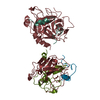
| ||||||||
|---|---|---|---|---|---|---|---|---|---|
| 1 | 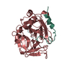
| ||||||||
| 2 | 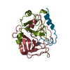
| ||||||||
| Unit cell |
| ||||||||
| Details | THERE ARE TWO INDEPENDENT COMPLEXES IN THE ASYMMETRIC UNIT: COMPLEX 1 EPSILON THROMBIN: CHAINS J, K, M FIBRINOPEPTIDE: CHAIN F COMPLEX 2 ALPHA THROMBIN: CHAINS L, H FIBRINOPEPTIDE: CHAIN N |
-Components
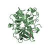
-Protein/peptide , 2 types, 4 molecules LJFN
| #1: Protein/peptide | Mass: 5735.240 Da / Num. of mol.: 2 / Source method: isolated from a natural source / Source: (natural) #3: Protein/peptide | Mass: 2349.497 Da / Num. of mol.: 2 / Fragment: RESIDUES 1 - 23 / Mutation: F308Y Source method: isolated from a genetically manipulated source References: UniProt: P02671 |
|---|
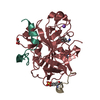
-Protein , 3 types, 3 molecules HKM
| #2: Protein | Mass: 29772.422 Da / Num. of mol.: 1 / Source method: isolated from a natural source / Source: (natural) |
|---|---|
| #4: Protein | Mass: 17525.346 Da / Num. of mol.: 1 / Source method: isolated from a natural source / Source: (natural) |
| #5: Protein | Mass: 12265.071 Da / Num. of mol.: 1 / Source method: isolated from a natural source / Source: (natural) |
-Non-polymers , 1 types, 276 molecules 
| #6: Water | ChemComp-HOH / Mass: 18.015 Da / Num. of mol.: 276 / Source method: isolated from a natural source / Formula: H2O |
|---|
-Details
| Has protein modification | Y |
|---|---|
| Sequence details | CHYMOTRYPSINOGEN NUMBERING (RATHER THAN SEQUENTIAL) SYSTEM IS USED, BASED ON THE TOPOLOGICAL ...CHYMOTRYPS |
-Experimental details
-Experiment
| Experiment | Method: X-RAY DIFFRACTION / Number of used crystals: 2 |
|---|
- Sample preparation
| Crystal | Density Matthews: 2.63 Å3/Da / Density % sol: 53.26 % | ||||||||||||||||||||||||||||||
|---|---|---|---|---|---|---|---|---|---|---|---|---|---|---|---|---|---|---|---|---|---|---|---|---|---|---|---|---|---|---|---|
| Crystal grow | pH: 7.5 Details: 2.0M AMMONIUM SULFATE 0.1M HEPES, PH 7.5 2.0% POLYETHYLENE GLYCOL 400 | ||||||||||||||||||||||||||||||
| Crystal grow | *PLUS pH: 6 / Method: vapor diffusion, hanging drop | ||||||||||||||||||||||||||||||
| Components of the solutions | *PLUS
|
-Data collection
| Diffraction | Mean temperature: 298 K |
|---|---|
| Diffraction source | Source: ROTATING ANODE / Type: RIGAKU / Wavelength: 1.5418 |
| Detector | Type: SIEMENS / Detector: AREA DETECTOR |
| Radiation | Monochromator: GRAPHITE(002) / Monochromatic (M) / Laue (L): M / Scattering type: x-ray |
| Radiation wavelength | Wavelength: 1.5418 Å / Relative weight: 1 |
| Reflection | Resolution: 2.35→7 Å / Num. obs: 18614 / % possible obs: 63 % / Observed criterion σ(I): 0 / Rmerge(I) obs: 0.109 |
| Reflection shell | *PLUS Highest resolution: 2.5 Å / Lowest resolution: 2.6 Å / % possible obs: 25 % |
- Processing
| Software |
| ||||||||||||||||||||||||||||||||||||||||||||||||||||||||||||
|---|---|---|---|---|---|---|---|---|---|---|---|---|---|---|---|---|---|---|---|---|---|---|---|---|---|---|---|---|---|---|---|---|---|---|---|---|---|---|---|---|---|---|---|---|---|---|---|---|---|---|---|---|---|---|---|---|---|---|---|---|---|
| Refinement | Method to determine structure: MOLECULAR REPLACEMENT / Resolution: 2.5→7 Å
| ||||||||||||||||||||||||||||||||||||||||||||||||||||||||||||
| Refinement step | Cycle: LAST / Resolution: 2.5→7 Å
| ||||||||||||||||||||||||||||||||||||||||||||||||||||||||||||
| Refine LS restraints |
| ||||||||||||||||||||||||||||||||||||||||||||||||||||||||||||
| Software | *PLUS Name: X-PLOR / Classification: refinement | ||||||||||||||||||||||||||||||||||||||||||||||||||||||||||||
| Refinement | *PLUS σ(I): 2 | ||||||||||||||||||||||||||||||||||||||||||||||||||||||||||||
| Solvent computation | *PLUS | ||||||||||||||||||||||||||||||||||||||||||||||||||||||||||||
| Displacement parameters | *PLUS | ||||||||||||||||||||||||||||||||||||||||||||||||||||||||||||
| Refine LS restraints | *PLUS
|
