 Movie
Movie Controller

[English] 日本語
 Yorodumi
- PDB-3dnm: Crystal Structure Hormone-Sensitive Lipase from a Metagenome Library -
+ Open data

- Basic information
| Entry | Database: PDB / ID: 3dnm | ||||||
|---|---|---|---|---|---|---|---|
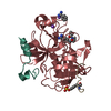
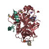
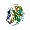
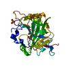
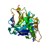
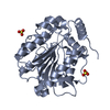
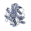
| Title | Crystal Structure Hormone-Sensitive Lipase from a Metagenome Library | ||||||
 Components Components | Esterase/lipase | ||||||
 Keywords Keywords | HYDROLASE / alpha/beta hydrolase fold | ||||||
| Function / homology |  Function and homology information Function and homology information | ||||||
| Biological species |  uncultured bacterium (environmental samples) uncultured bacterium (environmental samples) | ||||||
| Method |  X-RAY DIFFRACTION / SYNCHROTRON / MOLECULAR REPLACEMENT / Resolution: 2.8 Å X-RAY DIFFRACTION / SYNCHROTRON / MOLECULAR REPLACEMENT / Resolution: 2.8 Å | ||||||
 Authors Authors | Hwang, K.Y. / Nam, K.H. | ||||||
 Citation Citation | Journal: Proteins / Year: 2008 Title: Structural and functional analysis of a novel hormone-sensitive lipase from a metagenome library Authors: Nam, K.H. / Kim, M.-Y. / Kim, S.-J. / Priyadarshi, A. / Kwon, S.-T. / Koo, B.-S. / Yoon, S.-H. / Hwang, K.Y. | ||||||
| History |
|
- Structure visualization
| Structure viewer | Molecule: MolmilJmol/JSmol |
|---|
- Downloads & links
-Download
| PDBx/mmCIF format | 3dnm.cif.gz | 233.8 KB | Display | PDBx/mmCIF format |
|---|---|---|---|---|
| PDB format | pdb3dnm.ent.gz | 188 KB | Display | PDB format |
| PDBx/mmJSON format | 3dnm.json.gz | Tree view | PDBx/mmJSON format | |
| Others |  Other downloads Other downloads |
-Validation report
| Arichive directory | https://data.pdbj.org/pub/pdb/validation_reports/dn/3dnmftp://data.pdbj.org/pub/pdb/validation_reports/dn/3dnm | HTTPS FTP |
|---|
-Related structure data
| Related structure data |  1lzlS S: Starting model for refinement |
|---|---|
| Similar structure data |


-Links
 PDBj
PDBj


- Assembly
| Deposited unit | 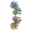
| ||||||||
|---|---|---|---|---|---|---|---|---|---|
| 1 | 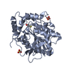
| ||||||||
| 2 | 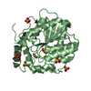
| ||||||||
| 3 | 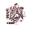
| ||||||||
| 4 | 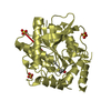
| ||||||||
| 5 | 
| ||||||||
| 6 | 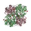
| ||||||||
| Unit cell |
| ||||||||
| Details | This protein works in monomer. But crystallographical data shows the dimer protein as reported in some related papers. The depositors consider that the dimer formation is crystallographical data. |
-Components
| #1: Protein | Mass: 36006.938 Da / Num. of mol.: 4 Source method: isolated from a genetically manipulated source Source: (gene. exp.) uncultured bacterium (environmental samples)Description: this protein was purified from Metagenome Library(soil uncultured bacteria) Gene: estE7 / Plasmid: pET21a / Production host: References: UniProt: Q0GMU1, Hydrolases; Acting on ester bonds; Carboxylic-ester hydrolases #2: Chemical | ChemComp-BME /   Mass: 78.133 Da / Num. of mol.: 4 / Source method: obtained synthetically / Formula: C2H6OS Mass: 78.133 Da / Num. of mol.: 4 / Source method: obtained synthetically / Formula: C2H6OS#3: Chemical | ChemComp-SO4 /   Mass: 96.063 Da / Num. of mol.: 14 / Source method: obtained synthetically / Formula: SO4 Mass: 96.063 Da / Num. of mol.: 14 / Source method: obtained synthetically / Formula: SO4#4: Water | ChemComp-HOH / |  Mass: 18.015 Da / Num. of mol.: 204 / Source method: isolated from a natural source / Formula: H2O Mass: 18.015 Da / Num. of mol.: 204 / Source method: isolated from a natural source / Formula: H2O |
|---|
-Experimental details
-Experiment
| Experiment | Method: X-RAY DIFFRACTION / Number of used crystals: 1 |
|---|
- Sample preparation
| Crystal | Density Matthews: 3.01 Å3/Da / Density % sol: 59.19 % |
|---|---|
| Crystal grow | Temperature: 295 K / Method: vapor diffusion, hanging drop / pH: 7 Details: 0.1M Bis-Tris propane, pH 7.0, 0.2M ammonium sulfate, 1M lithium sulfate, VAPOR DIFFUSION, HANGING DROP, temperature 295K |
-Data collection
| Diffraction | Mean temperature: 100 K |
|---|---|
| Diffraction source | Source: SYNCHROTRON / Site: PAL/PLS  / Beamline: 6C1 / Wavelength: 1.23 Å / Beamline: 6C1 / Wavelength: 1.23 Å |
| Detector | Type: ADSC QUANTUM 210 / Detector: CCD / Date: May 8, 2008 |
| Radiation | Monochromator: GRAPHITE / Protocol: SINGLE WAVELENGTH / Monochromatic (M) / Laue (L): M / Scattering type: x-ray |
| Radiation wavelength | Wavelength: 1.23 Å / Relative weight: 1 |
| Reflection | Resolution: 2.8→20 Å / Num. all: 47996 / Num. obs: 42217 / % possible obs: 88 % / Observed criterion σ(F): 2 / Observed criterion σ(I): 1 |
| Reflection shell | Resolution: 2.8→2.9 Å / % possible all: 96.9 |
- Processing
| Software |
| ||||||||||||||||||||||||||||||||||||||||||||||||||||||||||||||||||||||||||||||||
|---|---|---|---|---|---|---|---|---|---|---|---|---|---|---|---|---|---|---|---|---|---|---|---|---|---|---|---|---|---|---|---|---|---|---|---|---|---|---|---|---|---|---|---|---|---|---|---|---|---|---|---|---|---|---|---|---|---|---|---|---|---|---|---|---|---|---|---|---|---|---|---|---|---|---|---|---|---|---|---|---|---|
| Refinement | Method to determine structure: MOLECULAR REPLACEMENT Starting model: PDB ENTRY 1LZL Resolution: 2.8→20 Å / Cross valid method: THROUGHOUT / σ(F): 0 / Stereochemistry target values: Engh & Huber
| ||||||||||||||||||||||||||||||||||||||||||||||||||||||||||||||||||||||||||||||||
| Displacement parameters | Biso mean: 47.8071 Å2
| ||||||||||||||||||||||||||||||||||||||||||||||||||||||||||||||||||||||||||||||||
| Refinement step | Cycle: LAST / Resolution: 2.8→20 Å
| ||||||||||||||||||||||||||||||||||||||||||||||||||||||||||||||||||||||||||||||||
| Refine LS restraints |
|
