 Movie
Movie Controller

[English] 日本語
 Yorodumi
- PDB-1nro: CRYSTALLOGRAPHIC STRUCTURES OF THROMBIN COMPLEXED WITH THROMBIN R... -
+ Open data

- Basic information
| Entry | Database: PDB / ID: 1nro | ||||||
|---|---|---|---|---|---|---|---|
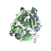
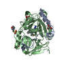
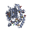
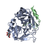
| Title | CRYSTALLOGRAPHIC STRUCTURES OF THROMBIN COMPLEXED WITH THROMBIN RECEPTOR PEPTIDES: EXISTENCE OF EXPECTED AND NOVEL BINDING MODES | ||||||
 Components Components |
| ||||||
 Keywords Keywords | SERINE PROTEINASE/RECEPTOR / SERINE PROTEINASE-RECEPTOR complex | ||||||
| Function / homology |  Function and homology information Function and homology informationnegative regulation of renin secretion into blood stream / trans-synaptic signaling by endocannabinoid, modulating synaptic transmission / platelet dense tubular network / establishment of synaptic specificity at neuromuscular junction / thrombin-activated receptor activity / connective tissue replacement involved in inflammatory response wound healing / regulation of interleukin-1 beta production / cell-cell junction maintenance / platelet dense granule organization / positive regulation of smooth muscle contraction ...negative regulation of renin secretion into blood stream / trans-synaptic signaling by endocannabinoid, modulating synaptic transmission / platelet dense tubular network / establishment of synaptic specificity at neuromuscular junction / thrombin-activated receptor activity / connective tissue replacement involved in inflammatory response wound healing / regulation of interleukin-1 beta production / cell-cell junction maintenance / platelet dense granule organization / positive regulation of smooth muscle contraction / positive regulation of calcium ion transport / : / thrombospondin receptor activity / thrombin / thrombin-activated receptor signaling pathway / Defective factor XII causes hereditary angioedema / negative regulation of glomerular filtration / negative regulation of astrocyte differentiation / regulation of blood coagulation / neutrophil-mediated killing of gram-negative bacterium / positive regulation of phospholipase C-activating G protein-coupled receptor signaling pathway / Defective F8 cleavage by thrombin / ligand-gated ion channel signaling pathway / Platelet Aggregation (Plug Formation) / positive regulation of Rho protein signal transduction / positive regulation of collagen biosynthetic process / negative regulation of platelet activation / negative regulation of blood coagulation / negative regulation of fibrinolysis / blood coagulation, fibrin clot formation / positive regulation of blood coagulation / positive regulation of vasoconstriction / anatomical structure morphogenesis / Transport of gamma-carboxylated protein precursors from the endoplasmic reticulum to the Golgi apparatus / : / G-protein alpha-subunit binding / Gamma-carboxylation of protein precursors / Removal of aminoterminal propeptides from gamma-carboxylated proteins / regulation of cytosolic calcium ion concentration / fibrinolysis / homeostasis of number of cells within a tissue / : / release of sequestered calcium ion into cytosol / negative regulation of proteolysis / negative regulation of cytokine production involved in inflammatory response / Regulation of Complement cascade / positive regulation of release of sequestered calcium ion into cytosol / acute-phase response / Cell surface interactions at the vascular wall / positive regulation of interleukin-8 production / Peptide ligand-binding receptors / growth factor activity / positive regulation of receptor signaling pathway via JAK-STAT / neuromuscular junction / lipopolysaccharide binding / platelet activation / positive regulation of protein localization to nucleus / regulation of synaptic plasticity / response to wounding / caveola / positive regulation of interleukin-6 production / Golgi lumen / Regulation of Insulin-like Growth Factor (IGF) transport and uptake by Insulin-like Growth Factor Binding Proteins (IGFBPs) / positive regulation of reactive oxygen species metabolic process / G protein-coupled receptor activity / blood coagulation / positive regulation of insulin secretion / late endosome / regulation of cell shape / antimicrobial humoral immune response mediated by antimicrobial peptide / heparin binding / G-protein beta-subunit binding / positive regulation of cytosolic calcium ion concentration / Thrombin signalling through proteinase activated receptors (PARs) / positive regulation of cell growth / response to lipopolysaccharide / blood microparticle / negative regulation of neuron apoptotic process / G alpha (q) signalling events / phospholipase C-activating G protein-coupled receptor signaling pathway / positive regulation of MAPK cascade / early endosome / positive regulation of ERK1 and ERK2 cascade / positive regulation of canonical NF-kappaB signal transduction / positive regulation of phosphatidylinositol 3-kinase/protein kinase B signal transduction / postsynaptic membrane / cell surface receptor signaling pathway / positive regulation of cell migration / positive regulation of apoptotic process / endoplasmic reticulum lumen / inflammatory response / G protein-coupled receptor signaling pathway / receptor ligand activity / negative regulation of cell population proliferation / serine-type endopeptidase activity / signaling receptor binding / calcium ion binding / positive regulation of cell population proliferation / positive regulation of DNA-templated transcription / Golgi apparatus Similarity search - Function | ||||||
| Biological species |  Homo sapiens (human) Homo sapiens (human) | ||||||
| Method |  X-RAY DIFFRACTION / Resolution: 3.1 Å X-RAY DIFFRACTION / Resolution: 3.1 Å | ||||||
 Authors Authors | Tulinsky, A. / Mathews, I.I. | ||||||
 Citation Citation | Journal: Biochemistry / Year: 1994 Title: Crystallographic structures of thrombin complexed with thrombin receptor peptides: existence of expected and novel binding modes. Authors: Mathews, I.I. / Padmanabhan, K.P. / Ganesh, V. / Tulinsky, A. / Ishii, M. / Chen, J. / Turck, C.W. / Coughlin, S.R. / Fenton 2nd., J.W. #1: Journal: J.Mol.Biol. / Year: 1991Title: Refined Structure of the Hirudin-Thrombin Complex Authors: Rydel, T.J. / Tulinsky, A. / Bode, W. / Huber, R. #2: Journal: J.Mol.Biol. / Year: 1991Title: Structure of the Hirugen and Hirulog 1 Complexes of Alpha-Thrombin Authors: Skrzypczak-Jankun, E. / Carperos, V.E. / Ravichandran, K.G. / Tulinsky, A. / Westbrook, M. / Maraganore, J.M. | ||||||
| History |
|
- Structure visualization
| Structure viewer | Molecule: MolmilJmol/JSmol |
|---|
- Downloads & links
-Download
| PDBx/mmCIF format | 1nro.cif.gz | 75.3 KB | Display | PDBx/mmCIF format |
|---|---|---|---|---|
| PDB format | pdb1nro.ent.gz | 55.2 KB | Display | PDB format |
| PDBx/mmJSON format | 1nro.json.gz | Tree view | PDBx/mmJSON format | |
| Others |  Other downloads Other downloads |
-Validation report
| Arichive directory | https://data.pdbj.org/pub/pdb/validation_reports/nr/1nroftp://data.pdbj.org/pub/pdb/validation_reports/nr/1nro | HTTPS FTP |
|---|
-Related structure data

-Links
 PDBj
PDBj


- Assembly
| Deposited unit | 
| ||||||||
|---|---|---|---|---|---|---|---|---|---|
| 1 |
| ||||||||
| Unit cell |
| ||||||||
| Atom site foot note | 1: CIS PROLINE - PRO H 37 2: A FEW SIDE CHAINS IN BOTH THROMBIN AND RECEPTOR PEPTIDE DO NOT HAVE WELL-DEFINED ELECTRON DENSITY. THESE ATOMS HAVE BEEN GIVEN OCCUPANCIES OF 0.01 IN THIS ENTRY. |
-Components
| #1: Protein/peptide | Mass: 4096.534 Da / Num. of mol.: 1 Source method: isolated from a genetically manipulated source Source: (gene. exp.) Homo sapiens (human) / References: UniProt: P00734, thrombin |
|---|---|
| #2: Protein | Mass: 29780.219 Da / Num. of mol.: 1 Source method: isolated from a genetically manipulated source Source: (gene. exp.) Homo sapiens (human) / References: UniProt: P00734, thrombin |
| #3: Protein/peptide | Mass: 3382.644 Da / Num. of mol.: 1 Source method: isolated from a genetically manipulated source Source: (gene. exp.) Homo sapiens (human) / References: UniProt: P25116 |
| #4: Water | ChemComp-HOH /  Mass: 18.015 Da / Num. of mol.: 117 / Source method: isolated from a natural source / Formula: H2O Mass: 18.015 Da / Num. of mol.: 117 / Source method: isolated from a natural source / Formula: H2O |
| Compound details | THROMBIN IS CLEAVED BETWEEN RESIDUES 15 AND 16. CHAIN INDICATOR *L* IS USED FOR RESIDUES 1H - 15 ...THROMBIN IS CLEAVED BETWEEN RESIDUES 15 AND 16. CHAIN INDICATOR *L* IS USED FOR RESIDUES 1H - 15 AND CHAIN INDICATOR *H* IS USED FOR RESIDUES 16 - 247. CHAIN INDICATOR *R* IS USED FOR NRP. |
| Has protein modification | Y |
-Experimental details
-Experiment
| Experiment | Method: X-RAY DIFFRACTION |
|---|
- Sample preparation
| Crystal | Density Matthews: 2.75 Å3/Da / Density % sol: 55.23 % | |||||||||||||||
|---|---|---|---|---|---|---|---|---|---|---|---|---|---|---|---|---|
| Crystal grow | *PLUS pH: 8.5 / Method: vapor diffusion, hanging drop | |||||||||||||||
| Components of the solutions | *PLUS
|
-Data collection
| Radiation | Scattering type: x-ray |
|---|---|
| Radiation wavelength | Relative weight: 1 |
| Reflection | *PLUS Highest resolution: 3.1 Å / % possible obs: 88 % / Rmerge F obs: 0.071 |
- Processing
| Software | Name: PROLSQ / Classification: refinement | ||||||||||||||||||||||||||||||||||||||||||||||||||||||||||||||||||||||||||||||||||||
|---|---|---|---|---|---|---|---|---|---|---|---|---|---|---|---|---|---|---|---|---|---|---|---|---|---|---|---|---|---|---|---|---|---|---|---|---|---|---|---|---|---|---|---|---|---|---|---|---|---|---|---|---|---|---|---|---|---|---|---|---|---|---|---|---|---|---|---|---|---|---|---|---|---|---|---|---|---|---|---|---|---|---|---|---|---|
| Refinement | Resolution: 3.1→7 Å / σ(F): 2 Details: A FEW SIDE CHAINS IN BOTH THROMBIN AND RECEPTOR PEPTIDE DO NOT HAVE WELL-DEFINED ELECTRON DENSITY. THESE ATOMS HAVE BEEN GIVEN OCCUPANCIES OF 0.01 IN THIS ENTRY.
| ||||||||||||||||||||||||||||||||||||||||||||||||||||||||||||||||||||||||||||||||||||
| Refinement step | Cycle: LAST / Resolution: 3.1→7 Å
| ||||||||||||||||||||||||||||||||||||||||||||||||||||||||||||||||||||||||||||||||||||
| Refine LS restraints |
| ||||||||||||||||||||||||||||||||||||||||||||||||||||||||||||||||||||||||||||||||||||
| Software | *PLUS Name: PROLSQ / Classification: refinement | ||||||||||||||||||||||||||||||||||||||||||||||||||||||||||||||||||||||||||||||||||||
| Refinement | *PLUS Rfactor obs: 0.171 | ||||||||||||||||||||||||||||||||||||||||||||||||||||||||||||||||||||||||||||||||||||
| Solvent computation | *PLUS | ||||||||||||||||||||||||||||||||||||||||||||||||||||||||||||||||||||||||||||||||||||
| Displacement parameters | *PLUS Biso mean: 29 Å2 |
