 Movie
Movie Controller

[English] 日本語
 Yorodumi
- PDB-2a45: Crystal structure of the complex between thrombin and the central... -
+ Open data

- Basic information
| Entry | Database: PDB / ID: 2a45 | |||||||||
|---|---|---|---|---|---|---|---|---|---|---|
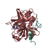
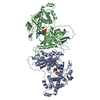
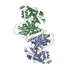
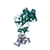
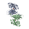
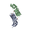
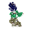
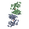
| Title | Crystal structure of the complex between thrombin and the central "E" region of fibrin | |||||||||
 Components Components |
| |||||||||
 Keywords Keywords | HYDROLASE/HYDROLASE INHIBITOR / THROMBIN / FIBRIN / FRAGMENT E / THROMBIN-FIBRIN COMPLEX / COILED COILS / DISULFIDE RINGS / BLOOD CLOTTING / HYDROLASE-HYDROLASE INHIBITOR COMPLEX | |||||||||
| Function / homology |  Function and homology information Function and homology informationblood coagulation, common pathway / induction of bacterial agglutination / fibrinogen complex / platelet alpha granule / Regulation of TLR by endogenous ligand / cellular response to leptin stimulus / MyD88 deficiency (TLR2/4) / positive regulation of heterotypic cell-cell adhesion / IRAK4 deficiency (TLR2/4) / plasminogen activation ...blood coagulation, common pathway / induction of bacterial agglutination / fibrinogen complex / platelet alpha granule / Regulation of TLR by endogenous ligand / cellular response to leptin stimulus / MyD88 deficiency (TLR2/4) / positive regulation of heterotypic cell-cell adhesion / IRAK4 deficiency (TLR2/4) / plasminogen activation / MyD88:MAL(TIRAP) cascade initiated on plasma membrane / extracellular matrix structural constituent / : / thrombospondin receptor activity / p130Cas linkage to MAPK signaling for integrins / thrombin / thrombin-activated receptor signaling pathway / Defective factor XII causes hereditary angioedema / positive regulation of peptide hormone secretion / negative regulation of astrocyte differentiation / regulation of blood coagulation / neutrophil-mediated killing of gram-negative bacterium / positive regulation of phospholipase C-activating G protein-coupled receptor signaling pathway / Defective F8 cleavage by thrombin / ligand-gated ion channel signaling pathway / Platelet Aggregation (Plug Formation) / GRB2:SOS provides linkage to MAPK signaling for Integrins / positive regulation of collagen biosynthetic process / positive regulation of exocytosis / negative regulation of platelet activation / negative regulation of blood coagulation / negative regulation of fibrinolysis / blood coagulation, fibrin clot formation / positive regulation of blood coagulation / protein polymerization / protein secretion / positive regulation of vasoconstriction / cellular response to interleukin-1 / Integrin cell surface interactions / Transport of gamma-carboxylated protein precursors from the endoplasmic reticulum to the Golgi apparatus / : / Gamma-carboxylation of protein precursors / negative regulation of endothelial cell apoptotic process / Removal of aminoterminal propeptides from gamma-carboxylated proteins / negative regulation of extrinsic apoptotic signaling pathway via death domain receptors / regulation of cytosolic calcium ion concentration / fibrinolysis / : / negative regulation of proteolysis / Integrin signaling / positive regulation of substrate adhesion-dependent cell spreading / negative regulation of cytokine production involved in inflammatory response / cell adhesion molecule binding / platelet alpha granule lumen / Regulation of Complement cascade / acute-phase response / cell-matrix adhesion / Cell surface interactions at the vascular wall / positive regulation of release of sequestered calcium ion into cytosol / Peptide ligand-binding receptors / growth factor activity / positive regulation of protein secretion / positive regulation of receptor signaling pathway via JAK-STAT / Post-translational protein phosphorylation / lipopolysaccharide binding / Signaling by high-kinase activity BRAF mutants / platelet activation / MAP2K and MAPK activation / response to calcium ion / response to wounding / positive regulation of protein localization to nucleus / Golgi lumen / platelet aggregation / Regulation of Insulin-like Growth Factor (IGF) transport and uptake by Insulin-like Growth Factor Binding Proteins (IGFBPs) / positive regulation of reactive oxygen species metabolic process / blood coagulation / positive regulation of insulin secretion / Signaling by RAF1 mutants / Signaling by moderate kinase activity BRAF mutants / Paradoxical activation of RAF signaling by kinase inactive BRAF / Signaling downstream of RAS mutants / Signaling by BRAF and RAF1 fusions / Platelet degranulation / regulation of cell shape / antimicrobial humoral immune response mediated by antimicrobial peptide / heparin binding / extracellular vesicle / protein-folding chaperone binding / Thrombin signalling through proteinase activated receptors (PARs) / extracellular matrix / positive regulation of cell growth / ER-Phagosome pathway / protein-containing complex assembly / blood microparticle / cell cortex / G alpha (q) signalling events / adaptive immune response / protein-macromolecule adaptor activity / positive regulation of ERK1 and ERK2 cascade / cell surface receptor signaling pathway Similarity search - Function | |||||||||
| Biological species |  Homo sapiens (human) Homo sapiens (human) | |||||||||
| Method |  X-RAY DIFFRACTION / MOLECULAR REPLACEMENT / Resolution: 3.65 Å X-RAY DIFFRACTION / MOLECULAR REPLACEMENT / Resolution: 3.65 Å | |||||||||
 Authors Authors | Pechik, I. / Madrazo, J. / Gilliland, G.L. / Medved, L. | |||||||||
 Citation Citation | Journal: Biochemistry / Year: 2006 Title: Structural basis for sequential cleavage of fibrinopeptides upon fibrin assembly. Authors: Pechik, I. / Yakovlev, S. / Mosesson, M.W. / Gilliland, G.L. / Medved, L. #1: Journal: Proc.Natl.Acad.Sci.USA / Year: 2004 Title: Crystal Structure of the Complex between Thrombin and the Central "E" Region of Fibrin Authors: Pechik, I. / Madrazo, J. / Mosesson, M.W. / Hernandez, I. / Gilliland, G.L. / Medved, L. | |||||||||
| History |
|
- Structure visualization
| Structure viewer | Molecule: MolmilJmol/JSmol |
|---|
- Downloads & links
-Download
| PDBx/mmCIF format | 2a45.cif.gz | 167.1 KB | Display | PDBx/mmCIF format |
|---|---|---|---|---|
| PDB format | pdb2a45.ent.gz | 125.8 KB | Display | PDB format |
| PDBx/mmJSON format | 2a45.json.gz | Tree view | PDBx/mmJSON format | |
| Others |  Other downloads Other downloads |
-Validation report
| Arichive directory | https://data.pdbj.org/pub/pdb/validation_reports/a4/2a45ftp://data.pdbj.org/pub/pdb/validation_reports/a4/2a45 | HTTPS FTP |
|---|
-Related structure data


-Links
 PDBj
PDBj


- Assembly
| Deposited unit | 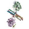
| ||||||||
|---|---|---|---|---|---|---|---|---|---|
| 1 |
| ||||||||
| Unit cell |
|
-Components
-Protein/peptide , 2 types, 4 molecules ADIL
| #1: Protein/peptide | Mass: 4096.534 Da / Num. of mol.: 2 / Source method: isolated from a natural source / Source: (natural) Homo sapiens (human) / References: UniProt: P00734, thrombin#5: Protein/peptide | Mass: 5103.651 Da / Num. of mol.: 2 / Source method: isolated from a natural source / Details: PROTEOLYTIC FRAGMENT / Source: (natural) Homo sapiens (human) / References: UniProt: P02679 |
|---|
-Protein , 3 types, 6 molecules BEGJHK
| #2: Protein | Mass: 29780.219 Da / Num. of mol.: 2 / Source method: isolated from a natural source / Source: (natural) Homo sapiens (human) / References: UniProt: P00734, thrombin#3: Protein | Mass: 6642.435 Da / Num. of mol.: 2 / Fragment: UNP P02671, residues 36-92 / Source method: isolated from a natural source / Details: PROTEOLYTIC FRAGMENT / Source: (natural) Homo sapiens (human) / References: UniProt: P02671#4: Protein | Mass: 9787.060 Da / Num. of mol.: 2 / Source method: isolated from a natural source / Details: PROTEOLYTIC FRAGMENT / Source: (natural) Homo sapiens (human) / References: UniProt: P02675 |
|---|
-Non-polymers , 2 types, 4 molecules 

| #6: Chemical |  Type: peptide-like, Peptide-like / Class: Inhibitor / Mass: 453.986 Da / Num. of mol.: 2 / Source method: obtained synthetically / Formula: C21H34ClN6O3 / References: D-Phe-Pro-Arg-CH2Cl Type: peptide-like, Peptide-like / Class: Inhibitor / Mass: 453.986 Da / Num. of mol.: 2 / Source method: obtained synthetically / Formula: C21H34ClN6O3 / References: D-Phe-Pro-Arg-CH2Cl#7: Chemical |  Mass: 94.971 Da / Num. of mol.: 2 / Source method: obtained synthetically / Formula: PO4 Mass: 94.971 Da / Num. of mol.: 2 / Source method: obtained synthetically / Formula: PO4 |
|---|
-Details
| Has protein modification | Y |
|---|---|
| Nonpolymer details | THE INHIBITOR IS COVALENTLY CONNECTED TO ACTIVE_SITE RESIDUES: 1) VIA A HEMIKETAL GROUP TO OG SER ...THE INHIBITOR IS COVALENTLY |
| Sequence details | HUMAN ALPHA THROMBIN COMPRISES AMINO ACID RESIDUES 1H TO 247 (CHYMOTRYPSIN NUMBERING). N-TERMINAL ...HUMAN ALPHA THROMBIN COMPRISES AMINO ACID RESIDUES 1H TO 247 (CHYMOTRYPS |
-Experimental details
-Experiment
| Experiment | Method: X-RAY DIFFRACTION / Number of used crystals: 1 |
|---|
- Sample preparation
| Crystal | Density Matthews: 2.89 Å3/Da / Density % sol: 55.58 % |
|---|---|
| Crystal grow | Temperature: 298 K / Method: microdialysis / pH: 7.9 Details: PEG 3500, AMMONIUM PHOSPHATE, TRIS, pH 7.90, MICRODIALYSIS, temperature 298K |
-Data collection
| Diffraction | Mean temperature: 100 K |
|---|---|
| Diffraction source | Source: ROTATING ANODE / Type: SIEMENS / Wavelength: 1.5418 |
| Detector | Type: MARRESEARCH / Detector: IMAGE PLATE / Details: COLLIMATOR |
| Radiation | Monochromator: GRAPHITE / Protocol: SINGLE WAVELENGTH / Monochromatic (M) / Laue (L): M / Scattering type: x-ray |
| Radiation wavelength | Wavelength: 1.5418 Å / Relative weight: 1 |
| Reflection | Resolution: 3.65→31.24 Å / Num. obs: 26890 / % possible obs: 96.3 % / Net I/σ(I): 3.4 |
| Reflection shell | Resolution: 3.65→3.78 Å / Mean I/σ(I) obs: 1.9 / % possible all: 87.6 |
- Processing
| Software |
| |||||||||||||||||||||||||||||||||
|---|---|---|---|---|---|---|---|---|---|---|---|---|---|---|---|---|---|---|---|---|---|---|---|---|---|---|---|---|---|---|---|---|---|---|
| Refinement | Method to determine structure: MOLECULAR REPLACEMENT Starting model: PDB entries 1PPB, 1JY2 Resolution: 3.65→19.7 Å / Cross valid method: THROUGHOUT / σ(F): 0 / Stereochemistry target values: ENGH & HUBER Details: DIFFRACTION DATA WERE COLLECTED FROM MEROHEDRALLY TWINNED CRYSTAL. THE PRESENCE OF 38.2% TWIN FRACTION CORRESPONDING TO -H -H-K -L TWINNING OPERATOR WAS TAKEN INTO ACCOUNT IN THE REFINEMENT. ...Details: DIFFRACTION DATA WERE COLLECTED FROM MEROHEDRALLY TWINNED CRYSTAL. THE PRESENCE OF 38.2% TWIN FRACTION CORRESPONDING TO -H -H-K -L TWINNING OPERATOR WAS TAKEN INTO ACCOUNT IN THE REFINEMENT.RESIDUES THAT ADDED THROUGH THE MODELING EFFORTS (26 THROUGH 28, CHAINS G, J) ARE INCLUDED IN THE COORDINATES FOR COMPLETENESS. THEY HAVE BEEN ASSIGNED OCCUPANCIES AND TEMPERATURE FACTORS OF 0.0, AND THEY HAVE NO VISIBLE/INTERPRETABLE ELECTRON DENSITY ASSOCIATED WITH THEIR LOCATIONS.
| |||||||||||||||||||||||||||||||||
| Solvent computation | Solvent model: FLAT MODEL | |||||||||||||||||||||||||||||||||
| Refinement step | Cycle: LAST / Resolution: 3.65→19.7 Å
| |||||||||||||||||||||||||||||||||
| Refine LS restraints |
|
