 Movie
Movie Controller

[English] 日本語
 Yorodumi
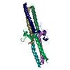
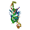
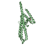
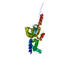
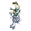
- PDB-1jy2: Crystal Structure of the Central Region of Bovine Fibrinogen (E5 ... -
+ Open data

- Basic information
| Entry | Database: PDB / ID: 1jy2 | ||||||
|---|---|---|---|---|---|---|---|
| Title | Crystal Structure of the Central Region of Bovine Fibrinogen (E5 fragment) at 1.4 Angstroms Resolution | ||||||
 Components Components |
| ||||||
 Keywords Keywords | BLOOD CLOTTING / fibrinogen / fragment E / disulfide bonds / asymmetry / coiled-coil / beta-sheet | ||||||
| Function / homology |  Function and homology information Function and homology informationblood coagulation, common pathway / fibrinogen complex / positive regulation of heterotypic cell-cell adhesion / blood coagulation, fibrin clot formation / protein polymerization / fibrinolysis / cell-matrix adhesion / platelet aggregation / adaptive immune response / protein-macromolecule adaptor activity ...blood coagulation, common pathway / fibrinogen complex / positive regulation of heterotypic cell-cell adhesion / blood coagulation, fibrin clot formation / protein polymerization / fibrinolysis / cell-matrix adhesion / platelet aggregation / adaptive immune response / protein-macromolecule adaptor activity / signaling receptor binding / innate immune response / : / metal ion binding Similarity search - Function | ||||||
| Biological species |  | ||||||
| Method |  X-RAY DIFFRACTION / SYNCHROTRON / MOLECULAR REPLACEMENT / Resolution: 1.4 Å X-RAY DIFFRACTION / SYNCHROTRON / MOLECULAR REPLACEMENT / Resolution: 1.4 Å | ||||||
 Authors Authors | Madrazo, J. / Brown, J.H. / Litvinovich, S. / Dominguez, R. / Yakovlev, S. / Medved, L. / Cohen, C. | ||||||
 Citation Citation | Journal: Proc.Natl.Acad.Sci.USA / Year: 2001 Title: Crystal structure of the central region of bovine fibrinogen (E5 fragment) at 1.4-A resolution. Authors: Madrazo, J. / Brown, J.H. / Litvinovich, S. / Dominguez, R. / Yakovlev, S. / Medved, L. / Cohen, C. #1: Journal: Proc.Natl.Acad.Sci.USA / Year: 2000Title: The Crystal Structure of Modified Bovine Fibrinogen Authors: Brown, J.H. / Volkmann, N. / Jun, G. / Henschen-Edman, A.H. / Cohen, C. | ||||||
| History |
| ||||||
| Remark 999 | SEQUENCE AN APPROPRIATE DATABASE MATCH WAS NOT AVAILABLE FOR FIBRINOGEN ALPHA CHAIN AT THE TIME OF PROCESSING. |
- Structure visualization
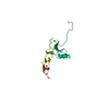
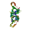
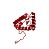
| Structure viewer | Molecule: MolmilJmol/JSmol |
|---|
- Downloads & links
-Download
| PDBx/mmCIF format | 1jy2.cif.gz | 73.9 KB | Display | PDBx/mmCIF format |
|---|---|---|---|---|
| PDB format | pdb1jy2.ent.gz | 54.9 KB | Display | PDB format |
| PDBx/mmJSON format | 1jy2.json.gz | Tree view | PDBx/mmJSON format | |
| Others |  Other downloads Other downloads |
-Validation report
| Arichive directory | https://data.pdbj.org/pub/pdb/validation_reports/jy/1jy2ftp://data.pdbj.org/pub/pdb/validation_reports/jy/1jy2 | HTTPS FTP |
|---|
-Related structure data

-Links
 PDBj
PDBj


- Assembly
| Deposited unit | 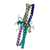
| ||||||||
|---|---|---|---|---|---|---|---|---|---|
| 1 |
| ||||||||
| Unit cell |
|
-Components
| #1: Protein | Mass: 6172.894 Da / Num. of mol.: 2 / Source method: isolated from a natural source / Details: Proteolytic fragment / Source: (natural) #2: Protein | Mass: 6253.134 Da / Num. of mol.: 2 / Source method: isolated from a natural source / Details: Proteolytic fragment / Source: (natural) #3: Protein/peptide | Mass: 5476.117 Da / Num. of mol.: 2 / Source method: isolated from a natural source / Details: Proteolytic fragment / Source: (natural) #4: Water | ChemComp-HOH / |  Mass: 18.015 Da / Num. of mol.: 318 / Source method: isolated from a natural source / Formula: H2O Mass: 18.015 Da / Num. of mol.: 318 / Source method: isolated from a natural source / Formula: H2OHas protein modification | Y | |
|---|
-Experimental details
-Experiment
| Experiment | Method: X-RAY DIFFRACTION / Number of used crystals: 1 |
|---|
- Sample preparation
| Crystal | Density Matthews: 2.22 Å3/Da / Density % sol: 44.5 % | |||||||||||||||||||||||||||||||||||||||||||||||||
|---|---|---|---|---|---|---|---|---|---|---|---|---|---|---|---|---|---|---|---|---|---|---|---|---|---|---|---|---|---|---|---|---|---|---|---|---|---|---|---|---|---|---|---|---|---|---|---|---|---|---|
| Crystal grow | Temperature: 295 K / Method: vapor diffusion / pH: 8 Details: PEG 3350, Calcium chloride, Tris, Dioxane, Sodium azide, pH 8.0, VAPOR DIFFUSION, temperature 295K | |||||||||||||||||||||||||||||||||||||||||||||||||
| Crystal grow | *PLUS Temperature: 4 ℃ / Method: vapor diffusion, hanging drop | |||||||||||||||||||||||||||||||||||||||||||||||||
| Components of the solutions | *PLUS
|
-Data collection
| Diffraction | Mean temperature: 100 K |
|---|---|
| Diffraction source | Source: SYNCHROTRON / Site: CHESS  / Beamline: A1 / Wavelength: 0.91 Å / Beamline: A1 / Wavelength: 0.91 Å |
| Detector | Type: ADSC QUANTUM 4 / Detector: CCD / Date: May 2, 1999 |
| Radiation | Protocol: SINGLE WAVELENGTH / Monochromatic (M) / Laue (L): M / Scattering type: x-ray |
| Radiation wavelength | Wavelength: 0.91 Å / Relative weight: 1 |
| Reflection | Resolution: 1.4→100 Å / Num. all: 61149 / Num. obs: 61149 / % possible obs: 99.2 % / Redundancy: 5.8 % / Rsym value: 0.058 |
| Reflection shell | Resolution: 1.4→1.45 Å / Rsym value: 0.339 / % possible all: 93.1 |
| Reflection | *PLUS Num. measured all: 359638 / Rmerge(I) obs: 0.058 |
| Reflection shell | *PLUS Rmerge(I) obs: 0.334 |
- Processing
| Software |
| |||||||||||||||||||||||||
|---|---|---|---|---|---|---|---|---|---|---|---|---|---|---|---|---|---|---|---|---|---|---|---|---|---|---|
| Refinement | Method to determine structure: MOLECULAR REPLACEMENT Starting model: 1.6 Angstrom resolution structure of bovine E5 in the orthorhombic space group Resolution: 1.4→100 Å / σ(F): 0
| |||||||||||||||||||||||||
| Displacement parameters | Biso mean: 28.8 Å2
| |||||||||||||||||||||||||
| Refine analyze |
| |||||||||||||||||||||||||
| Refinement step | Cycle: LAST / Resolution: 1.4→100 Å
| |||||||||||||||||||||||||
| Refine LS restraints |
| |||||||||||||||||||||||||
| LS refinement shell |
| |||||||||||||||||||||||||
| Software | *PLUS Name: CNS / Version: 1 / Classification: refinement | |||||||||||||||||||||||||
| Refinement | *PLUS Highest resolution: 1.4 Å / Lowest resolution: 100 Å / σ(F): 0 / % reflection Rfree: 5 % / Rfactor obs: 0.216 | |||||||||||||||||||||||||
| Solvent computation | *PLUS | |||||||||||||||||||||||||
| Displacement parameters | *PLUS Biso mean: 28.8 Å2 | |||||||||||||||||||||||||
| Refine LS restraints | *PLUS
| |||||||||||||||||||||||||
| LS refinement shell | *PLUS Rfactor Rfree: 0.411 / Rfactor Rwork: 0.398 |
