 Movie
Movie Controller


+ Open data

- Basic information
| Entry | Database: PDB / ID: 2cav | ||||||
|---|---|---|---|---|---|---|---|
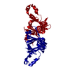
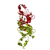
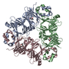
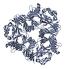
| Title | CANAVALIN FROM JACK BEAN | ||||||
 Components Components | PROTEIN (CANAVALIN) | ||||||
 Keywords Keywords | PLANT PROTEIN / VICILIN / 7S SEED PROTEIN / DOMAIN DUPLICATION / SWISS ROLL | ||||||
| Function / homology |  Function and homology information Function and homology informationnutrient reservoir activity / protein-containing complex / identical protein binding Similarity search - Function | ||||||
| Biological species |   Canavalia ensiformis (jack bean) Canavalia ensiformis (jack bean) | ||||||
| Method |  X-RAY DIFFRACTION / MIR / Resolution: 2 Å X-RAY DIFFRACTION / MIR / Resolution: 2 Å | ||||||
 Authors Authors | Ko, T.-P. / Day, J. / Macpherson, A. | ||||||
 Citation Citation | Journal: Acta Crystallogr.,Sect.D / Year: 2000 Title: The refined structure of canavalin from jack bean in two crystal forms at 2.1 and 2.0 A resolution. Authors: Ko, T.P. / Day, J. / McPherson, A. #1: Journal: Plant Physiol. / Year: 1993Title: The Three-Dimensional Structure of Canavalin from Jack Bean (Canavalia Ensiformis) Authors: Ko, T.-P. / Ng, J.D. / Macpherson, A. | ||||||
| History |
|
- Structure visualization
| Structure viewer | Molecule: MolmilJmol/JSmol |
|---|
- Downloads & links
-Download
| PDBx/mmCIF format | 2cav.cif.gz | 84.5 KB | Display | PDBx/mmCIF format |
|---|---|---|---|---|
| PDB format | pdb2cav.ent.gz | 64.4 KB | Display | PDB format |
| PDBx/mmJSON format | 2cav.json.gz | Tree view | PDBx/mmJSON format | |
| Others |  Other downloads Other downloads |
-Validation report
| Arichive directory | https://data.pdbj.org/pub/pdb/validation_reports/ca/2cavftp://data.pdbj.org/pub/pdb/validation_reports/ca/2cav | HTTPS FTP |
|---|
-Related structure data


-Links
 PDBj
PDBj- Assembly
| Deposited unit | 
| ||||||||
|---|---|---|---|---|---|---|---|---|---|
| 1 | 
| ||||||||
| Unit cell |
|
-Components
| #1: Protein | Mass: 50383.469 Da / Num. of mol.: 1 / Source method: isolated from a natural source / Details: TREATED WITH LIMITED DIGESTION BY TRYPSIN / Source: (natural) Canavalia ensiformis (jack bean) / Cellular location: PROTEIN BODY / Organ: SEED / Tissue: COTYLEDON / References: UniProt: P50477 |
|---|---|
| #2: Water | ChemComp-HOH /  Mass: 18.015 Da / Num. of mol.: 164 / Source method: isolated from a natural source / Formula: H2O Mass: 18.015 Da / Num. of mol.: 164 / Source method: isolated from a natural source / Formula: H2O |
-Experimental details
-Experiment
| Experiment | Method: X-RAY DIFFRACTION / Number of used crystals: 1 |
|---|
- Sample preparation
| Crystal | Density Matthews: 2.5 Å3/Da / Density % sol: 48 % / Description: REFERENCE TO PDB1CAV | ||||||||||||||||||||||||
|---|---|---|---|---|---|---|---|---|---|---|---|---|---|---|---|---|---|---|---|---|---|---|---|---|---|
| Crystal grow | Method: vapor diffusion / pH: 6.8 Details: 30 - 40 MG/ML PROTEIN SOLUTION WAS PREPARED BY DISSOLVING 4-TIME RECRYSTALLIZED CANAVALIN IN DISTILLED WATER PLUS TRACE NH4OH. RESERVOIR SOLUTION CONTAINED 2.0 % NACL IN 50 MM PHOSPHATE ...Details: 30 - 40 MG/ML PROTEIN SOLUTION WAS PREPARED BY DISSOLVING 4-TIME RECRYSTALLIZED CANAVALIN IN DISTILLED WATER PLUS TRACE NH4OH. RESERVOIR SOLUTION CONTAINED 2.0 % NACL IN 50 MM PHOSPHATE BUFFER AT PH 6.8. CRYSTALS WERE OBTAINED BY MIXING PROTEIN AND RESERVOIR SOLUTION IN SITTING DROPS FOLLOWED BY VAPOR DIFFUSION AGAINST THE RESERVOIR., vapor diffusion | ||||||||||||||||||||||||
| Crystal | *PLUS | ||||||||||||||||||||||||
| Crystal grow | *PLUS Temperature: 277-281 K / Method: vapor diffusion | ||||||||||||||||||||||||
| Components of the solutions | *PLUS
|
-Data collection
| Diffraction | Mean temperature: 290 K |
|---|---|
| Diffraction source | Source: ROTATING ANODE / Type: RIGAKU RU200 / Wavelength: 1.5418 |
| Detector | Type: SDMS / Detector: AREA DETECTOR / Date: Aug 1, 1994 |
| Radiation | Monochromator: GRAPHITE CRYSTAL / Protocol: SINGLE WAVELENGTH / Monochromatic (M) / Laue (L): M / Scattering type: x-ray |
| Radiation wavelength | Wavelength: 1.5418 Å / Relative weight: 1 |
| Reflection | Resolution: 2→40 Å / Num. obs: 31848 / % possible obs: 99.7 % / Redundancy: 10.6 % / Rmerge(I) obs: 0.101 / Net I/σ(I): 24 |
| Reflection shell | Resolution: 2→2.15 Å / Redundancy: 4.7 % / Rmerge(I) obs: 0.312 / Mean I/σ(I) obs: 3.4 / % possible all: 99.6 |
| Reflection | *PLUS Num. measured all: 337907 |
| Reflection shell | *PLUS % possible obs: 99.6 % / Num. unique obs: 6302 / Num. measured obs: 29912 |
- Processing
| Software |
| ||||||||||||||||||||||||||||||||||||||||||||||||||||||||||||||||||||||||||||||||
|---|---|---|---|---|---|---|---|---|---|---|---|---|---|---|---|---|---|---|---|---|---|---|---|---|---|---|---|---|---|---|---|---|---|---|---|---|---|---|---|---|---|---|---|---|---|---|---|---|---|---|---|---|---|---|---|---|---|---|---|---|---|---|---|---|---|---|---|---|---|---|---|---|---|---|---|---|---|---|---|---|---|
| Refinement | Method to determine structure: MIR / Resolution: 2→40 Å / Data cutoff high absF: 10000000 / Data cutoff low absF: 0.001 / Isotropic thermal model: RESTRAINED / Cross valid method: THROUGHOUT / σ(F): 2
| ||||||||||||||||||||||||||||||||||||||||||||||||||||||||||||||||||||||||||||||||
| Displacement parameters | Biso mean: 37.1 Å2 | ||||||||||||||||||||||||||||||||||||||||||||||||||||||||||||||||||||||||||||||||
| Refine analyze |
| ||||||||||||||||||||||||||||||||||||||||||||||||||||||||||||||||||||||||||||||||
| Refinement step | Cycle: LAST / Resolution: 2→40 Å
| ||||||||||||||||||||||||||||||||||||||||||||||||||||||||||||||||||||||||||||||||
| Refine LS restraints |
| ||||||||||||||||||||||||||||||||||||||||||||||||||||||||||||||||||||||||||||||||
| LS refinement shell | Resolution: 2→2.07 Å / Total num. of bins used: 10
| ||||||||||||||||||||||||||||||||||||||||||||||||||||||||||||||||||||||||||||||||
| Xplor file |
| ||||||||||||||||||||||||||||||||||||||||||||||||||||||||||||||||||||||||||||||||
| Software | *PLUS Name: X-PLOR / Version: 3.54 / Classification: refinement | ||||||||||||||||||||||||||||||||||||||||||||||||||||||||||||||||||||||||||||||||
| Refinement | *PLUS σ(F): 2 / % reflection Rfree: 8 % | ||||||||||||||||||||||||||||||||||||||||||||||||||||||||||||||||||||||||||||||||
| Solvent computation | *PLUS | ||||||||||||||||||||||||||||||||||||||||||||||||||||||||||||||||||||||||||||||||
| Displacement parameters | *PLUS Biso mean: 37.1 Å2 | ||||||||||||||||||||||||||||||||||||||||||||||||||||||||||||||||||||||||||||||||
| Refine LS restraints | *PLUS
| ||||||||||||||||||||||||||||||||||||||||||||||||||||||||||||||||||||||||||||||||
| LS refinement shell | *PLUS Rfactor Rfree: 0.37 / % reflection Rfree: 6.9 % / Rfactor Rwork: 0.333 / Num. reflection obs: 2647 / Rfactor obs: 0.333 |
