Mass: 18.015 Da / Num. of mol.: 82 / Source method: isolated from a natural source / Formula: H2O
Has ligand of interest
Y
-
Experimental details
-
Experiment
Experiment
Method: X-RAY DIFFRACTION / Number of used crystals: 1
-
Sample preparation
Crystal
Density Matthews: 2.43 Å3/Da / Density % sol: 49 % / Description: elongated regtangular prisms
Crystal grow
Temperature: 298 K / Method: vapor diffusion, sitting drop / pH: 7.5 Details: Crystals were grown by sitting drop vapor diffusion from 18% PEG 3350 in 0.1 M HEPES reservoirs. The drops were equal amounts of a 40 mg/ml solution of precanavalin (not treated with any ...Details: Crystals were grown by sitting drop vapor diffusion from 18% PEG 3350 in 0.1 M HEPES reservoirs. The drops were equal amounts of a 40 mg/ml solution of precanavalin (not treated with any exogenous protease) in water. Crystals grew only after six to eight weeks at room temperature. PH range: 7.0 - 7.8
-
Data collection
Diffraction
Mean temperature: 173 K / Serial crystal experiment: N
Diffraction source
Source: SYNCHROTRON / Site: ALS / Beamline: 8.3.1 / Wavelength: 1 Å
Detector
Type: DECTRIS PILATUS3 R CdTe 300K / Detector: PIXEL / Date: Jun 15, 2018
Radiation
Protocol: SINGLE WAVELENGTH / Monochromatic (M) / Laue (L): M / Scattering type: x-ray
Resolution: 2.8→30 Å / Cor.coef. Fo:Fc: 0.948 / Cor.coef. Fo:Fc free: 0.904 / SU B: 51.145 / SU ML: 0.386 / Cross valid method: THROUGHOUT / σ(F): 0 / ESU R Free: 0.438 / Stereochemistry target values: MAXIMUM LIKELIHOOD Details: HYDROGENS HAVE BEEN ADDED IN THE RIDING POSITIONS U VALUES : WITH TLS ADDED
Rfactor
Num. reflection
% reflection
Selection details
Rfree
0.252
1442
5.1 %
RANDOM
Rwork
0.1933
-
-
-
obs
0.1962
27012
78.86 %
-
Solvent computation
Ion probe radii: 0.8 Å / Shrinkage radii: 0.8 Å / VDW probe radii: 1.2 Å / Solvent model: MASK
In the structure databanks used in Yorodumi, some data are registered as the other names, "COVID-19 virus" and "2019-nCoV". Here are the details of the virus and the list of structure data.
Jan 31, 2019. EMDB accession codes are about to change! (news from PDBe EMDB page)
EMDB accession codes are about to change! (news from PDBe EMDB page)
The allocation of 4 digits for EMDB accession codes will soon come to an end. Whilst these codes will remain in use, new EMDB accession codes will include an additional digit and will expand incrementally as the available range of codes is exhausted. The current 4-digit format prefixed with “EMD-” (i.e. EMD-XXXX) will advance to a 5-digit format (i.e. EMD-XXXXX), and so on. It is currently estimated that the 4-digit codes will be depleted around Spring 2019, at which point the 5-digit format will come into force.
The EM Navigator/Yorodumi systems omit the EMD- prefix.
Related info.:Q: What is EMD? / ID/Accession-code notation in Yorodumi/EM Navigator
Yorodumi is a browser for structure data from EMDB, PDB, SASBDB, etc.
This page is also the successor to EM Navigator detail page, and also detail information page/front-end page for Omokage search.
The word "yorodu" (or yorozu) is an old Japanese word meaning "ten thousand". "mi" (miru) is to see.
Related info.:EMDB / PDB / SASBDB / Comparison of 3 databanks / Yorodumi Search / Aug 31, 2016. New EM Navigator & Yorodumi / Yorodumi Papers / Jmol/JSmol / Function and homology information / Changes in new EM Navigator and Yorodumi
 Movie
Movie Controller


 Open data
 Basic information
 Components
 Keywords
 Function and homology information
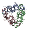
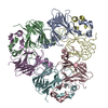
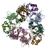
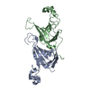
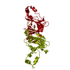
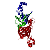
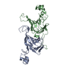
 Canavalia ensiformis (jack bean)
 X-RAY DIFFRACTION /
  Authors
 Citation
 Structure visualization
 Downloads & links
 Other downloads
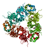


 PDBj
PDBj Assembly
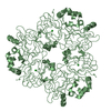

 Mass: 122.121 Da / Num. of mol.: 3 / Source method: obtained synthetically / Formula: C7H6O2 / Feature type: SUBJECT OF INVESTIGATION
 Mass: 18.015 Da / Num. of mol.: 82 / Source method: isolated from a natural source / Formula: H2O
 Sample preparation
 / Beamline: 8.3.1 / Wavelength: 1 Å
 Processing