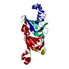
 Movie
Movie Controller

[English] 日本語
 Yorodumi
- PDB-6v7g: Binding of Benzoic Acid and Anions Within the Cupin Domains of th... -
+ Open data

- Basic information
| Entry | Database: PDB / ID: 6v7g | |||||||||
|---|---|---|---|---|---|---|---|---|---|---|
| Title | Binding of Benzoic Acid and Anions Within the Cupin Domains of the Vicillin Protein Canavalin from Jack Bean (canavalia ensiformis): Crystal Structures | |||||||||
 Components Components | Canavalin | |||||||||
 Keywords Keywords | PLANT PROTEIN / plant proteins / ligands / salicylic acid / enzymes | |||||||||
| Function / homology |  Function and homology information Function and homology informationnutrient reservoir activity / protein-containing complex / identical protein binding Similarity search - Function | |||||||||
| Biological species |   Canavalia ensiformis (jack bean) Canavalia ensiformis (jack bean) | |||||||||
| Method |  X-RAY DIFFRACTION / SYNCHROTRON / MOLECULAR REPLACEMENT / Resolution: 1.4 Å X-RAY DIFFRACTION / SYNCHROTRON / MOLECULAR REPLACEMENT / Resolution: 1.4 Å | |||||||||
 Authors Authors | McPherson, A. | |||||||||
 Citation Citation | Journal: Biochem.Biophys.Res.Commun. / Year: 2020 Title: Binding of benzoic acid and anions within the cupin domains of the vicilin protein canavalin from jack bean (Canavalia ensiformis): Crystal structures. Authors: McPherson, A. | |||||||||
| History |
|
- Structure visualization
| Structure viewer | Molecule: MolmilJmol/JSmol |
|---|
- Downloads & links
-Download
| PDBx/mmCIF format | 6v7g.cif.gz | 225.4 KB | Display | PDBx/mmCIF format |
|---|---|---|---|---|
| PDB format | pdb6v7g.ent.gz | 184.6 KB | Display | PDB format |
| PDBx/mmJSON format | 6v7g.json.gz | Tree view | PDBx/mmJSON format | |
| Others |  Other downloads Other downloads |
-Validation report
| Arichive directory | https://data.pdbj.org/pub/pdb/validation_reports/v7/6v7gftp://data.pdbj.org/pub/pdb/validation_reports/v7/6v7g | HTTPS FTP |
|---|
-Related structure data
| Related structure data | 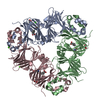 6v7jC 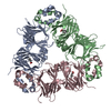 6v7lC  6cb4 S: Starting model for refinement C: citing same article ( |
|---|---|
| Similar structure data |


-Links
 PDBj
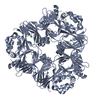
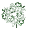
PDBj- Assembly
| Deposited unit | 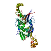
| ||||||||
|---|---|---|---|---|---|---|---|---|---|
| 1 | 
| ||||||||
| Unit cell |
|
-Components
| #1: Protein | Mass: 50431.469 Da / Num. of mol.: 1 / Source method: isolated from a natural source / Source: (natural) Canavalia ensiformis (jack bean) / References: UniProt: P50477 |
|---|---|
| #2: Chemical | ChemComp-ACT /   Mass: 59.044 Da / Num. of mol.: 1 / Source method: obtained synthetically / Formula: C2H3O2 Mass: 59.044 Da / Num. of mol.: 1 / Source method: obtained synthetically / Formula: C2H3O2 |
| #3: Chemical | ChemComp-BEZ /   Mass: 122.121 Da / Num. of mol.: 1 / Source method: obtained synthetically / Formula: C7H6O2 / Feature type: SUBJECT OF INVESTIGATION Mass: 122.121 Da / Num. of mol.: 1 / Source method: obtained synthetically / Formula: C7H6O2 / Feature type: SUBJECT OF INVESTIGATION |
| #4: Chemical | ChemComp-CIT /   Mass: 192.124 Da / Num. of mol.: 1 / Source method: obtained synthetically / Formula: C6H8O7 Mass: 192.124 Da / Num. of mol.: 1 / Source method: obtained synthetically / Formula: C6H8O7 |
| #5: Water | ChemComp-HOH /  Mass: 18.015 Da / Num. of mol.: 273 / Source method: isolated from a natural source / Formula: H2O Mass: 18.015 Da / Num. of mol.: 273 / Source method: isolated from a natural source / Formula: H2O |
| Has ligand of interest | Y |
-Experimental details
-Experiment
| Experiment | Method: X-RAY DIFFRACTION / Number of used crystals: 1 |
|---|
- Sample preparation
| Crystal | Density Matthews: 2.3 Å3/Da / Density % sol: 46 % / Description: short hexagonal prisms |
|---|---|
| Crystal grow | Temperature: 298 K / Method: vapor diffusion, sitting drop / pH: 6 Details: Vapor diffusion in sitting drop Cryschem plates. Reservoirs were 1.0 M sodium citrate titrated with acetic acid to pH6.0. Drops were initially equal amounts of the reservoir solution with a ...Details: Vapor diffusion in sitting drop Cryschem plates. Reservoirs were 1.0 M sodium citrate titrated with acetic acid to pH6.0. Drops were initially equal amounts of the reservoir solution with a protein stock solution of 30 mg/ml canavalin in water with a trace of ammonium hydroxide. At room temperature crystallization time was about three weeks. PH range: 5.8 - 6.5 |
-Data collection
| Diffraction | Mean temperature: 173 K / Serial crystal experiment: N |
|---|---|
| Diffraction source | Source: SYNCHROTRON / Site: ALS  / Beamline: 8.3.1 / Wavelength: 1 Å / Beamline: 8.3.1 / Wavelength: 1 Å |
| Detector | Type: DECTRIS PILATUS3 R CdTe 300K / Detector: PIXEL / Date: Jun 15, 2018 |
| Radiation | Protocol: SINGLE WAVELENGTH / Monochromatic (M) / Laue (L): M / Scattering type: x-ray |
| Radiation wavelength | Wavelength: 1 Å / Relative weight: 1 |
| Reflection | Resolution: 1.4→109 Å / Num. obs: 86853 / % possible obs: 99.6 % / Redundancy: 61 % / Biso Wilson estimate: 14.6 Å2 / CC1/2: 0.99 / Rmerge(I) obs: 0.258 / Rpim(I) all: 0.029 / Rrim(I) all: 0.26 / Rsym value: 0.248 / Net I/σ(I): 18.7 |
| Reflection shell | Resolution: 1.4→1.43 Å / Redundancy: 11.8 % / Mean I/σ(I) obs: 0.7 / Num. unique obs: 3978 / CC1/2: 0.182 / % possible all: 92.8 |
- Processing
| Software |
| ||||||||||||||||||||||||||||||||||||||||||||||||||||||||||||
|---|---|---|---|---|---|---|---|---|---|---|---|---|---|---|---|---|---|---|---|---|---|---|---|---|---|---|---|---|---|---|---|---|---|---|---|---|---|---|---|---|---|---|---|---|---|---|---|---|---|---|---|---|---|---|---|---|---|---|---|---|---|
| Refinement | Method to determine structure: MOLECULAR REPLACEMENT Starting model: 6CB4 6cb4 Resolution: 1.4→109 Å / Cor.coef. Fo:Fc: 0.978 / Cor.coef. Fo:Fc free: 0.967 / SU B: 2.607 / SU ML: 0.046 / Cross valid method: THROUGHOUT / σ(F): 0 / ESU R: 0.049 / ESU R Free: 0.053 Details: HYDROGENS HAVE BEEN ADDED IN THE RIDING POSITIONS U VALUES : WITH TLS ADDED
| ||||||||||||||||||||||||||||||||||||||||||||||||||||||||||||
| Solvent computation | Ion probe radii: 0.8 Å / Shrinkage radii: 0.8 Å / VDW probe radii: 1.2 Å | ||||||||||||||||||||||||||||||||||||||||||||||||||||||||||||
| Displacement parameters | Biso max: 120.09 Å2 / Biso mean: 28.707 Å2 / Biso min: 15.02 Å2
| ||||||||||||||||||||||||||||||||||||||||||||||||||||||||||||
| Refinement step | Cycle: final / Resolution: 1.4→109 Å
| ||||||||||||||||||||||||||||||||||||||||||||||||||||||||||||
| Refine LS restraints |
| ||||||||||||||||||||||||||||||||||||||||||||||||||||||||||||
| LS refinement shell | Resolution: 1.4→1.436 Å / Rfactor Rfree error: 0 / Total num. of bins used: 20
| ||||||||||||||||||||||||||||||||||||||||||||||||||||||||||||
| Refinement TLS params. | Method: refined / Origin x: 21.2274 Å / Origin y: 151.639 Å / Origin z: 1.1447 Å
|
