 Movie
Movie Controller


+ Open data

- Basic information
| Entry | Database: PDB / ID: 2eaa | ||||||
|---|---|---|---|---|---|---|---|
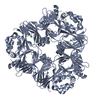
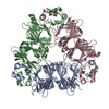
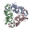
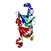
| Title | Crystal Structure of Adzuki Bean 7S Globulin-3 | ||||||
 Components Components | 7S globulin-3 | ||||||
 Keywords Keywords | PLANT PROTEIN / BETA BARREL / CUPIN SUPERFAMILY | ||||||
| Function / homology |  Function and homology information Function and homology information | ||||||
| Biological species |  Vigna angularis (adzuki bean) Vigna angularis (adzuki bean) | ||||||
| Method |  X-RAY DIFFRACTION / SYNCHROTRON / MOLECULAR REPLACEMENT / Resolution: 2.25 Å X-RAY DIFFRACTION / SYNCHROTRON / MOLECULAR REPLACEMENT / Resolution: 2.25 Å | ||||||
 Authors Authors | Fukuda, T. / Mikami, B. / Utsumi, S. | ||||||
 Citation Citation | Journal: J.Agric.Food Chem. / Year: 2008 Title: Characterization and crystallography of recombinant 7S globulins of Adzuki bean and structure-function relationships with 7S globulins of various crops. Authors: Fukuda, T. / Maruyama, N. / Salleh, M.R. / Mikami, B. / Utsumi, S. | ||||||
| History |
|
- Structure visualization
| Structure viewer | Molecule: MolmilJmol/JSmol |
|---|
- Downloads & links
-Download
| PDBx/mmCIF format | 2eaa.cif.gz | 248.8 KB | Display | PDBx/mmCIF format |
|---|---|---|---|---|
| PDB format | pdb2eaa.ent.gz | 199.3 KB | Display | PDB format |
| PDBx/mmJSON format | 2eaa.json.gz | Tree view | PDBx/mmJSON format | |
| Others |  Other downloads Other downloads |
-Validation report
| Arichive directory | https://data.pdbj.org/pub/pdb/validation_reports/ea/2eaaftp://data.pdbj.org/pub/pdb/validation_reports/ea/2eaa | HTTPS FTP |
|---|
-Related structure data
| Related structure data | 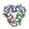 2ea7SC S: Starting model for refinement C: citing same article ( |
|---|---|
| Similar structure data |


-Links
 PDBj
PDBj

- Assembly
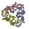
| Deposited unit | 
| ||||||||
|---|---|---|---|---|---|---|---|---|---|
| 1 |
| ||||||||
| Unit cell |
|
-Components
| #1: Protein | Mass: 49823.512 Da / Num. of mol.: 3 Source method: isolated from a genetically manipulated source Source: (gene. exp.) Vigna angularis (adzuki bean) / Gene: 7S-3 / Plasmid: pEAdzuki7S3 / Production host:  #2: Chemical |   Mass: 40.078 Da / Num. of mol.: 3 / Source method: obtained synthetically / Formula: Ca Mass: 40.078 Da / Num. of mol.: 3 / Source method: obtained synthetically / Formula: Ca#3: Chemical |   Mass: 192.124 Da / Num. of mol.: 3 / Source method: obtained synthetically / Formula: C6H8O7 Mass: 192.124 Da / Num. of mol.: 3 / Source method: obtained synthetically / Formula: C6H8O7#4: Chemical |   Mass: 60.052 Da / Num. of mol.: 3 / Source method: obtained synthetically / Formula: C2H4O2 Mass: 60.052 Da / Num. of mol.: 3 / Source method: obtained synthetically / Formula: C2H4O2#5: Water | ChemComp-HOH / |  Mass: 18.015 Da / Num. of mol.: 481 / Source method: isolated from a natural source / Formula: H2O Mass: 18.015 Da / Num. of mol.: 481 / Source method: isolated from a natural source / Formula: H2O |
|---|
-Experimental details
-Experiment
| Experiment | Method: X-RAY DIFFRACTION / Number of used crystals: 1 |
|---|
- Sample preparation
| Crystal | Density Matthews: 2.41 Å3/Da / Density % sol: 48.94 % |
|---|---|
| Crystal grow | Temperature: 293 K / Method: vapor diffusion, hanging drop / pH: 5.6 Details: 30% MPD, 0.2M ammonium acetate, 0.1M Sodium citrate, 2.0M ammonium sulfate, pH 5.6, VAPOR DIFFUSION, HANGING DROP, temperature 293.0K |
-Data collection
| Diffraction | Mean temperature: 100 K |
|---|---|
| Diffraction source | Source: SYNCHROTRON / Site: SPring-8  / Beamline: BL38B1 / Wavelength: 1 Å / Beamline: BL38B1 / Wavelength: 1 Å |
| Detector | Type: RIGAKU RAXIS V / Detector: IMAGE PLATE / Date: Oct 4, 2006 / Details: mirror |
| Radiation | Monochromator: Si 111 CHANNEL / Protocol: SINGLE WAVELENGTH / Monochromatic (M) / Laue (L): M / Scattering type: x-ray |
| Radiation wavelength | Wavelength: 1 Å / Relative weight: 1 |
| Reflection | Resolution: 2.25→50 Å / Num. all: 69062 / Num. obs: 69062 / % possible obs: 100 % / Observed criterion σ(I): 1 / Redundancy: 5.5 % / Biso Wilson estimate: 24.2 Å2 / Rmerge(I) obs: 0.056 / Net I/σ(I): 19 |
| Reflection shell | Resolution: 2.25→2.33 Å / Redundancy: 5.3 % / Rmerge(I) obs: 0.415 / Num. unique all: 6784 / % possible all: 100 |
- Processing
| Software |
| ||||||||||||||||||||||||||||||||||||
|---|---|---|---|---|---|---|---|---|---|---|---|---|---|---|---|---|---|---|---|---|---|---|---|---|---|---|---|---|---|---|---|---|---|---|---|---|---|
| Refinement | Method to determine structure: MOLECULAR REPLACEMENT Starting model: PDB ENTRY 2EA7 Resolution: 2.25→15 Å / Rfactor Rfree error: 0.003 / Data cutoff high absF: 2656502.27 / Data cutoff low absF: 0 / Isotropic thermal model: RESTRAINED / Cross valid method: THROUGHOUT / σ(F): 0 / Stereochemistry target values: Engh & Huber
| ||||||||||||||||||||||||||||||||||||
| Solvent computation | Solvent model: FLAT MODEL / Bsol: 44.4143 Å2 / ksol: 0.351261 e/Å3 | ||||||||||||||||||||||||||||||||||||
| Displacement parameters | Biso mean: 39.5 Å2
| ||||||||||||||||||||||||||||||||||||
| Refine analyze |
| ||||||||||||||||||||||||||||||||||||
| Refinement step | Cycle: LAST / Resolution: 2.25→15 Å
| ||||||||||||||||||||||||||||||||||||
| Refine LS restraints |
| ||||||||||||||||||||||||||||||||||||
| LS refinement shell | Resolution: 2.25→2.39 Å / Rfactor Rfree error: 0.01 / Total num. of bins used: 6
| ||||||||||||||||||||||||||||||||||||
| Xplor file |
|
