 Movie
Movie Controller

[English] 日本語
 Yorodumi
- PDB-1ipj: CRYSTAL STRUCTURES OF RECOMBINANT AND NATIVE SOYBEAN BETA-CONGLYC... -
+ Open data

- Basic information
| Entry | Database: PDB / ID: 1ipj | ||||||
|---|---|---|---|---|---|---|---|
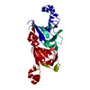
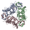
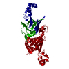
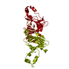
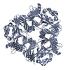
| Title | CRYSTAL STRUCTURES OF RECOMBINANT AND NATIVE SOYBEAN BETA-CONGLYCININ BETA HOMOTRIMERS COMPLEXES WITH N-ACETYL-D-GLUCOSAMINE | ||||||
 Components Components | BETA-CONGLYCININ, BETA CHAIN | ||||||
 Keywords Keywords | SUGAR BINDING PROTEIN / soybean / storage protein / vicilin | ||||||
| Function / homology |  Function and homology information Function and homology informationaleurone grain / protein storage vacuole / nutrient reservoir activity / endoplasmic reticulum Similarity search - Function | ||||||
| Biological species |  | ||||||
| Method |  X-RAY DIFFRACTION / MOLECULAR REPLACEMENT / Resolution: 2.7 Å X-RAY DIFFRACTION / MOLECULAR REPLACEMENT / Resolution: 2.7 Å | ||||||
 Authors Authors | Maruyama, N. / Adachi, M. / Takahashi, K. / Yagasaki, K. / Kohno, M. / Takenaka, Y. / Okuda, E. / Nakagawa, S. / Mikami, B. / Utsumi, S. | ||||||
 Citation Citation | Journal: Eur.J.Biochem. / Year: 2001 Title: Crystal structures of recombinant and native soybean beta-conglycinin beta homotrimers. Authors: Maruyama, N. / Adachi, M. / Takahashi, K. / Yagasaki, K. / Kohno, M. / Takenaka, Y. / Okuda, E. / Nakagawa, S. / Mikami, B. / Utsumi, S. | ||||||
| History |
|
- Structure visualization
| Structure viewer | Molecule: MolmilJmol/JSmol |
|---|
- Downloads & links
-Download
| PDBx/mmCIF format | 1ipj.cif.gz | 226.4 KB | Display | PDBx/mmCIF format |
|---|---|---|---|---|
| PDB format | pdb1ipj.ent.gz | 182.3 KB | Display | PDB format |
| PDBx/mmJSON format | 1ipj.json.gz | Tree view | PDBx/mmJSON format | |
| Others |  Other downloads Other downloads |
-Validation report
| Arichive directory | https://data.pdbj.org/pub/pdb/validation_reports/ip/1ipjftp://data.pdbj.org/pub/pdb/validation_reports/ip/1ipj | HTTPS FTP |
|---|
-Related structure data


-Links
 PDBj
PDBj- Assembly
| Deposited unit | 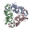
| ||||||||
|---|---|---|---|---|---|---|---|---|---|
| 1 |
| ||||||||
| Unit cell |
|
-Components
| #1: Protein | Mass: 47965.461 Da / Num. of mol.: 3 / Source method: isolated from a natural source / Source: (natural) #2: Sugar |   Type: D-saccharide, beta linking / Mass: 221.208 Da / Num. of mol.: 3 Type: D-saccharide, beta linking / Mass: 221.208 Da / Num. of mol.: 3Source method: isolated from a genetically manipulated source Formula: C8H15NO6 Has protein modification | Y | |
|---|
-Experimental details
-Experiment
| Experiment | Method: X-RAY DIFFRACTION / Number of used crystals: 1 |
|---|
- Sample preparation
| Crystal | Density Matthews: 2.48 Å3/Da / Density % sol: 50.49 % | |||||||||||||||||||||||||||||||||||||||||||||||||||||||||||||||||||||||||||||||||||||||||||
|---|---|---|---|---|---|---|---|---|---|---|---|---|---|---|---|---|---|---|---|---|---|---|---|---|---|---|---|---|---|---|---|---|---|---|---|---|---|---|---|---|---|---|---|---|---|---|---|---|---|---|---|---|---|---|---|---|---|---|---|---|---|---|---|---|---|---|---|---|---|---|---|---|---|---|---|---|---|---|---|---|---|---|---|---|---|---|---|---|---|---|---|---|
| Crystal grow | Temperature: 277 K / Method: vapor diffusion, hanging drop / pH: 4.8 Details: PEG6000, citrate buffer, pH 4.8, VAPOR DIFFUSION, HANGING DROP, temperature 277K | |||||||||||||||||||||||||||||||||||||||||||||||||||||||||||||||||||||||||||||||||||||||||||
| Crystal grow | *PLUS Temperature: 4 ℃ / pH: 5.5 | |||||||||||||||||||||||||||||||||||||||||||||||||||||||||||||||||||||||||||||||||||||||||||
| Components of the solutions | *PLUS
|
-Data collection
| Diffraction | Mean temperature: 293 K |
|---|---|
| Diffraction source | Source: ROTATING ANODE / Type: MACSCIENCE / Wavelength: 1.5418 Å |
| Detector | Type: SIEMENS HI-STAR / Detector: AREA DETECTOR / Date: Jul 4, 1998 |
| Radiation | Protocol: SINGLE WAVELENGTH / Monochromatic (M) / Laue (L): M / Scattering type: x-ray |
| Radiation wavelength | Wavelength: 1.5418 Å / Relative weight: 1 |
| Reflection | Highest resolution: 2.8 Å / Num. all: 33726 / Num. obs: 72174 / % possible obs: 94.37 % / Redundancy: 2.14 % / Rsym value: 0.055 |
| Reflection | *PLUS Highest resolution: 2.7 Å / Num. obs: 33726 / Num. measured all: 72174 / Rmerge(I) obs: 0.051 |
- Processing
| Software |
| |||||||||||||||||||||
|---|---|---|---|---|---|---|---|---|---|---|---|---|---|---|---|---|---|---|---|---|---|---|
| Refinement | Method to determine structure: MOLECULAR REPLACEMENT / Resolution: 2.7→7 Å / σ(F): 2 / σ(I): 0
| |||||||||||||||||||||
| Refinement step | Cycle: LAST / Resolution: 2.7→7 Å
| |||||||||||||||||||||
| Refinement | *PLUS Rfactor obs: 0.202 | |||||||||||||||||||||
| Solvent computation | *PLUS | |||||||||||||||||||||
| Displacement parameters | *PLUS | |||||||||||||||||||||
| Refine LS restraints | *PLUS
|
