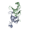
 Movie
Movie Controller


+ Open data

- Basic information
| Entry | Database: PDB / ID: 1dgr | ||||||
|---|---|---|---|---|---|---|---|
| Title | Refined crystal structure of canavalin from jack bean | ||||||
 Components Components | (CANAVALIN) x 3 | ||||||
 Keywords Keywords | PLANT PROTEIN / DUPLICATED DOMAINS BETA BARREL HELICAL LOOP | ||||||
| Function / homology |  Function and homology information Function and homology informationnutrient reservoir activity / protein-containing complex / identical protein binding Similarity search - Function | ||||||
| Biological species |   Canavalia ensiformis (jack bean) Canavalia ensiformis (jack bean) | ||||||
| Method |  X-RAY DIFFRACTION / Resolution: 2.6 Å X-RAY DIFFRACTION / Resolution: 2.6 Å | ||||||
 Authors Authors | Ko, T.-P. / McPherson, A. | ||||||
 Citation Citation | Journal: Acta Crystallogr.,Sect.D / Year: 2001 Title: X-ray diffraction and atomic force microscopy analysis of twinned crystals: rhombohedral canavalin. Authors: Ko, T.P. / Kuznetsov, Y.G. / Malkin, A.J. / Day, J. / McPherson, A. #1: Journal: Acta Crystallogr.,Sect.D / Year: 1993Title: Determination of three crystal structures of canavalin by molecular replacement Authors: Ko, T.-P. / Ng, J.D. / Day, J. / Greenwood, A. / McPherson, A. | ||||||
| History |
|
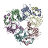
- Structure visualization
| Structure viewer | Molecule: MolmilJmol/JSmol |
|---|
- Downloads & links
-Download
| PDBx/mmCIF format | 1dgr.cif.gz | 212.8 KB | Display | PDBx/mmCIF format |
|---|---|---|---|---|
| PDB format | pdb1dgr.ent.gz | 172.8 KB | Display | PDB format |
| PDBx/mmJSON format | 1dgr.json.gz | Tree view | PDBx/mmJSON format | |
| Others |  Other downloads Other downloads |
-Validation report
| Arichive directory | https://data.pdbj.org/pub/pdb/validation_reports/dg/1dgrftp://data.pdbj.org/pub/pdb/validation_reports/dg/1dgr | HTTPS FTP |
|---|
-Related structure data


-Links
 PDBj
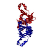
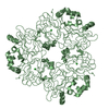
PDBj- Assembly
| Deposited unit | 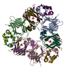
| ||||||||
|---|---|---|---|---|---|---|---|---|---|
| 1 |
| ||||||||
| Unit cell |
| ||||||||
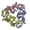
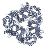
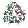
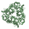
| Details | The trimeric assembly contains three identical subunits of chains A,X,Y; B,V,W; and C,N,M. |
-Components
| #1: Protein | Mass: 20640.342 Da / Num. of mol.: 3 / Fragment: RESIDUES 46-223 / Source method: isolated from a natural source Details: LEGUME STORAGE PROTEIN; LIMITED DIGESTION BY TRYPSIN Source: (natural) Canavalia ensiformis (jack bean) / Organ: SEED / Tissue: COTYLEDON / References: UniProt: P50477#2: Protein | Mass: 9073.182 Da / Num. of mol.: 3 / Fragment: RESIDUES 246-324 / Source method: isolated from a natural source Details: LEGUME STORAGE PROTEIN; LIMITED DIGESTION BY TRYPSIN Source: (natural) Canavalia ensiformis (jack bean) / Organ: SEED / Tissue: COTYLEDON / References: UniProt: P50477#3: Protein | Mass: 10269.446 Da / Num. of mol.: 3 / Fragment: RESIDUES 331-423 / Source method: isolated from a natural source Details: LEGUME STORAGE PROTEIN; LIMITED DIGESTION BY TRYPSIN Source: (natural) Canavalia ensiformis (jack bean) / Organ: SEED / Tissue: COTYLEDON / References: UniProt: P50477#4: Chemical |   Mass: 94.971 Da / Num. of mol.: 3 / Source method: obtained synthetically / Formula: PO4 Mass: 94.971 Da / Num. of mol.: 3 / Source method: obtained synthetically / Formula: PO4#5: Water | ChemComp-HOH / |  Mass: 18.015 Da / Num. of mol.: 168 / Source method: isolated from a natural source / Formula: H2O Mass: 18.015 Da / Num. of mol.: 168 / Source method: isolated from a natural source / Formula: H2O |
|---|
-Experimental details
-Experiment
| Experiment | Method: X-RAY DIFFRACTION / Number of used crystals: 1 |
|---|
- Sample preparation
| Crystal | Density Matthews: 2.85 Å3/Da / Density % sol: 56.87 % |
|---|---|
| Crystal grow | Temperature: 277 K / Method: vapor diffusion, sitting drop / pH: 6.8 Details: Dulbeccos phosphate buffered saline, ammonium hydroxide, pH 6.8, VAPOR DIFFUSION, SITTING DROP, temperature 277K |
| Crystal grow | *PLUS Details: Koszelak, S., (1995) Biophys. J., 69, 13. |
-Data collection
| Diffraction | Mean temperature: 298 K |
|---|---|
| Diffraction source | Source: ROTATING ANODE / Type: RIGAKU RU200 / Wavelength: 1.5418 |
| Detector | Type: SDMS / Detector: AREA DETECTOR / Date: Jan 23, 1989 |
| Radiation | Protocol: SINGLE WAVELENGTH / Monochromatic (M) / Laue (L): M / Scattering type: x-ray |
| Radiation wavelength | Wavelength: 1.5418 Å / Relative weight: 1 |
| Reflection | Resolution: 2.6→200 Å / Num. all: 42478 / Num. obs: 37938 / % possible obs: 89.3 % / Redundancy: 3.7 % / Biso Wilson estimate: 23.5 Å2 / Rmerge(I) obs: 0.0548 / Net I/σ(I): 21.3 |
| Reflection shell | Resolution: 2.6→2.8 Å / Redundancy: 2.58 % / Rmerge(I) obs: 0.1744 / Num. unique all: 7058 / % possible all: 83 |
- Processing
| Software |
| |||||||||||||||||||||||||
|---|---|---|---|---|---|---|---|---|---|---|---|---|---|---|---|---|---|---|---|---|---|---|---|---|---|---|
| Refinement | Resolution: 2.6→200 Å / σ(F): 3 / σ(I): 0 / Stereochemistry target values: Engh & Huber / Details: Strong NCS restraints were used.
| |||||||||||||||||||||||||
| Refinement step | Cycle: LAST / Resolution: 2.6→200 Å
| |||||||||||||||||||||||||
| Refine LS restraints |
| |||||||||||||||||||||||||
| Software | *PLUS Name: CNS / Classification: refinement | |||||||||||||||||||||||||
| Refinement | *PLUS Lowest resolution: 500 Å / σ(F): 3 / Rfactor obs: 0.181 / Rfactor Rwork: 0.18 / % reflection Rfree: 8 % | |||||||||||||||||||||||||
| Solvent computation | *PLUS | |||||||||||||||||||||||||
| Displacement parameters | *PLUS | |||||||||||||||||||||||||
| Refine LS restraints | *PLUS
| |||||||||||||||||||||||||
| LS refinement shell | *PLUS Highest resolution: 2.6 Å / Lowest resolution: 2.76 Å / Rfactor Rfree: 0.263 / Rfactor Rwork: 0.23 / Num. reflection obs: 2283 |
