 Movie
Movie Controller

[English] 日本語
 Yorodumi
- PDB-1cav: THE THREE-DIMENSIONAL STRUCTURE OF CANAVALIN FROM JACK BEAN (CANA... -
+ Open data

- Basic information
| Entry | Database: PDB / ID: 1cav | ||||||
|---|---|---|---|---|---|---|---|
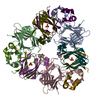
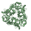
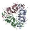
| Title | THE THREE-DIMENSIONAL STRUCTURE OF CANAVALIN FROM JACK BEAN (CANAVALIA ENSIFORMIS) | ||||||
 Components Components | (CANAVALIN) x 2 | ||||||
 Keywords Keywords | SEED STORAGE PROTEIN | ||||||
| Function / homology |  Function and homology information Function and homology informationnutrient reservoir activity / protein-containing complex / identical protein binding Similarity search - Function | ||||||
| Biological species |   Canavalia ensiformis (jack bean) Canavalia ensiformis (jack bean) | ||||||
| Method |  X-RAY DIFFRACTION / Resolution: 2.6 Å X-RAY DIFFRACTION / Resolution: 2.6 Å | ||||||
 Authors Authors | Ko, T-P. / Ng, J.D. / McPherson, A. | ||||||
 Citation Citation | Journal: Acta Crystallogr.,Sect.D / Year: 1993 Title: Determination of three crystal structures of canavalin by molecular replacement. Authors: Ko, T.P. / Ng, J.D. / Day, J. / Greenwood, A. / McPherson, A. #1: Journal: Plant Physiol. / Year: 1993 Title: The three-dimensional structure of canavalin from jack bean (Canavalia ensiformis). Authors: Ko, T.-P. / Ng, J.D. / Day, J. / Greenwood, A. / McPherson, A. | ||||||
| History |
|
- Structure visualization
| Structure viewer | Molecule: MolmilJmol/JSmol |
|---|
- Downloads & links
-Download
| PDBx/mmCIF format | 1cav.cif.gz | 84.3 KB | Display | PDBx/mmCIF format |
|---|---|---|---|---|
| PDB format | pdb1cav.ent.gz | 64.4 KB | Display | PDB format |
| PDBx/mmJSON format | 1cav.json.gz | Tree view | PDBx/mmJSON format | |
| Others |  Other downloads Other downloads |
-Validation report
| Arichive directory | https://data.pdbj.org/pub/pdb/validation_reports/ca/1cavftp://data.pdbj.org/pub/pdb/validation_reports/ca/1cav | HTTPS FTP |
|---|
-Related structure data


-Links
 PDBj
PDBj- Assembly
| Deposited unit | 
| ||||||||
|---|---|---|---|---|---|---|---|---|---|
| 1 | 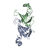
| ||||||||
| Unit cell |
|
-Components
| #1: Protein | Mass: 20968.727 Da / Num. of mol.: 1 Source method: isolated from a genetically manipulated source Source: (gene. exp.) Canavalia ensiformis (jack bean) / References: UniProt: P50477 |
|---|---|
| #2: Protein | Mass: 20641.021 Da / Num. of mol.: 1 Source method: isolated from a genetically manipulated source Source: (gene. exp.) Canavalia ensiformis (jack bean) / References: UniProt: P50477 |
-Experimental details
-Experiment
| Experiment | Method: X-RAY DIFFRACTION |
|---|
- Sample preparation
| Crystal | Density Matthews: 2.86 Å3/Da / Density % sol: 56.97 % | ||||||||||||||||||||||||
|---|---|---|---|---|---|---|---|---|---|---|---|---|---|---|---|---|---|---|---|---|---|---|---|---|---|
| Crystal grow | *PLUS Temperature: 4, 8 ℃ / pH: 6.8 / Method: vapor diffusion | ||||||||||||||||||||||||
| Components of the solutions | *PLUS
|
-Data collection
| Radiation | Scattering type: x-ray |
|---|---|
| Radiation wavelength | Relative weight: 1 |
- Processing
| Software | Name: TNT / Classification: refinement | ||||||||||||||||||||||||||||||
|---|---|---|---|---|---|---|---|---|---|---|---|---|---|---|---|---|---|---|---|---|---|---|---|---|---|---|---|---|---|---|---|
| Refinement | Resolution: 2.6→8 Å / σ(I): 3 /
| ||||||||||||||||||||||||||||||
| Refinement step | Cycle: LAST / Resolution: 2.6→8 Å
| ||||||||||||||||||||||||||||||
| Refine LS restraints |
| ||||||||||||||||||||||||||||||
| Refine LS restraints | *PLUS
|
