 Movie
Movie Controller

[English] 日本語
 Yorodumi
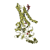
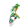
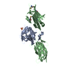
- PDB-3zqe: PrgI-SipD from Salmonella typhimurium in complex with deoxycholate -
+ Open data

- Basic information
| Entry | Database: PDB / ID: 3zqe | ||||||
|---|---|---|---|---|---|---|---|
| Title | PrgI-SipD from Salmonella typhimurium in complex with deoxycholate | ||||||
 Components Components | PROTEIN PRGI, CELL INVASION PROTEIN SIPD | ||||||
 Keywords Keywords | CELL INVASION / CELL INVASION COMPLEX / TYPE III SECRETION / T3SS / TIP COMPLEX / HOST PATHOGEN INTERACTION / BACTERIAL PATHOGENESIS / BILE SALT | ||||||
| Function / homology |  Function and homology information Function and homology informationtype III protein secretion system complex / protein secretion by the type III secretion system / cell surface / extracellular region / identical protein binding Similarity search - Function | ||||||
| Biological species |  SALMONELLA ENTERICA SUBSP. ENTERICA SEROVAR TYPHIMURIUM (bacteria) SALMONELLA ENTERICA SUBSP. ENTERICA SEROVAR TYPHIMURIUM (bacteria) | ||||||
| Method |  X-RAY DIFFRACTION / SYNCHROTRON / MOLECULAR REPLACEMENT / Resolution: 2.19 Å X-RAY DIFFRACTION / SYNCHROTRON / MOLECULAR REPLACEMENT / Resolution: 2.19 Å | ||||||
 Authors Authors | Lunelli, M. / Kolbe, M. | ||||||
 Citation Citation | Journal: Plos Pathog. / Year: 2011 Title: Crystal Structure of Prgi-Sipd: Insight Into a Secretion Competent State of the Type Three Secretion System Needle Tip and its Interaction with Host Ligands Authors: Lunelli, M. / Hurwitz, R. / Lambers, J. / Kolbe, M. | ||||||
| History |
|
- Structure visualization
| Structure viewer | Molecule: MolmilJmol/JSmol |
|---|
- Downloads & links
-Download
| PDBx/mmCIF format | 3zqe.cif.gz | 116.6 KB | Display | PDBx/mmCIF format |
|---|---|---|---|---|
| PDB format | pdb3zqe.ent.gz | 89.1 KB | Display | PDB format |
| PDBx/mmJSON format | 3zqe.json.gz | Tree view | PDBx/mmJSON format | |
| Others |  Other downloads Other downloads |
-Validation report
| Arichive directory | https://data.pdbj.org/pub/pdb/validation_reports/zq/3zqeftp://data.pdbj.org/pub/pdb/validation_reports/zq/3zqe | HTTPS FTP |
|---|
-Related structure data
| Related structure data | 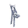 2ym0C 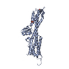 2ym9C  3zqbSC C: citing same article ( S: Starting model for refinement |
|---|---|
| Similar structure data |


-Links
 PDBj
PDBj- Assembly
| Deposited unit | 
| ||||||||
|---|---|---|---|---|---|---|---|---|---|
| 1 | 
| ||||||||
| 2 | 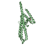
| ||||||||
| Unit cell |
| ||||||||
| Noncrystallographic symmetry (NCS) | NCS oper: (Code: given Matrix: (-0.6858, -0.6888, 0.235), Vector: |
-Components
| #1: Protein | Mass: 33158.875 Da / Num. of mol.: 2 / Fragment: PRGI FUSED WITH SIPD RESIDUES 127-343 Source method: isolated from a genetically manipulated source Details: GGSGG LINK INSERTED BETWEEN PRGI AND SIPD Source: (gene. exp.) SALMONELLA ENTERICA SUBSP. ENTERICA SEROVAR TYPHIMURIUM (bacteria)Strain: SL1344 / Production host: #2: Chemical |   Mass: 92.094 Da / Num. of mol.: 2 / Source method: obtained synthetically / Formula: C3H8O3 Mass: 92.094 Da / Num. of mol.: 2 / Source method: obtained synthetically / Formula: C3H8O3#3: Chemical | ChemComp-DXC / ( | 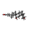  Mass: 392.572 Da / Num. of mol.: 1 / Source method: obtained synthetically / Formula: C24H40O4 / Comment: detergent*YM Mass: 392.572 Da / Num. of mol.: 1 / Source method: obtained synthetically / Formula: C24H40O4 / Comment: detergent*YM#4: Water | ChemComp-HOH / |  Mass: 18.015 Da / Num. of mol.: 97 / Source method: isolated from a natural source / Formula: H2O Mass: 18.015 Da / Num. of mol.: 97 / Source method: isolated from a natural source / Formula: H2ONonpolymer details | (3ALPHA,5BETA,12ALPHA)-3,12-DIHYDROXYC | Sequence details | RESIDUES GSH AT THE N-TERMINUS REMAINING AFTER HIS-TAG CLEAVAGE. GGSGG LINKER INSERTED BETWEEN PRGI ...RESIDUES GSH AT THE N-TERMINUS REMAINING AFTER HIS-TAG CLEAVAGE. GGSGG LINKER INSERTED BETWEEN PRGI AND SIPD127-343. | |
|---|
-Experimental details
-Experiment
| Experiment | Method: X-RAY DIFFRACTION / Number of used crystals: 1 |
|---|
- Sample preparation
| Crystal | Density Matthews: 2.67 Å3/Da / Density % sol: 53.9 % / Description: NONE |
|---|---|
| Crystal grow | Details: PROTEIN WAS MIXED WITH SOLUTION CONTAINING 0.026 M NAH2PO4 AND 1.26 M K2HPO4. OBTAINED CRYSTALS WERE SOAKED FOR 72 HOURS IN SAME SOLUTION CONTAINING 0.01 M DEOXYCHOLATE. |
-Data collection
| Diffraction | Mean temperature: 100 K |
|---|---|
| Diffraction source | Source: SYNCHROTRON / Site: BESSY  / Beamline: 14.2 / Wavelength: 0.91841 / Beamline: 14.2 / Wavelength: 0.91841 |
| Detector | Type: MARMOSAIC 225 mm CCD / Detector: CCD / Date: May 4, 2010 / Details: MIRRORS |
| Radiation | Monochromator: SI-111 CRYSTAL / Protocol: SINGLE WAVELENGTH / Monochromatic (M) / Laue (L): M / Scattering type: x-ray |
| Radiation wavelength | Wavelength: 0.91841 Å / Relative weight: 1 |
| Reflection | Resolution: 2.19→40 Å / Num. obs: 34906 / % possible obs: 95.4 % / Observed criterion σ(I): -3 / Redundancy: 5.8 % / Biso Wilson estimate: 46.6 Å2 / Rmerge(I) obs: 0.07 / Net I/σ(I): 16.02 |
| Reflection shell | Resolution: 2.19→2.26 Å / Redundancy: 5.2 % / Rmerge(I) obs: 0.48 / Mean I/σ(I) obs: 4.73 / % possible all: 84.4 |
- Processing
| Software |
| ||||||||||||||||||||||||||||||||||||||||||||||||||||||||||||||||||||||||||||||||
|---|---|---|---|---|---|---|---|---|---|---|---|---|---|---|---|---|---|---|---|---|---|---|---|---|---|---|---|---|---|---|---|---|---|---|---|---|---|---|---|---|---|---|---|---|---|---|---|---|---|---|---|---|---|---|---|---|---|---|---|---|---|---|---|---|---|---|---|---|---|---|---|---|---|---|---|---|---|---|---|---|---|
| Refinement | Method to determine structure: MOLECULAR REPLACEMENT Starting model: PDB ENTRY 3ZQB Resolution: 2.19→36.48 Å / Rfactor Rfree error: 0.007 / Data cutoff high absF: 3170360.76 / Data cutoff low absF: 0 / Isotropic thermal model: RESTRAINED / Cross valid method: THROUGHOUT / σ(F): 0 / Stereochemistry target values: MAXIMUM LIKELIHOOD Details: BULK SOLVENT MODEL USED. THE CHAINS WERE RENUMBERED TO BE CONSISTENT WITH NUMBERING OF THE BIOLOGICALLY ACTIVE MOLECULES FOR WHICH THERE ARE EXISTING PDB AND UNIPROT ENTRIES.
| ||||||||||||||||||||||||||||||||||||||||||||||||||||||||||||||||||||||||||||||||
| Solvent computation | Solvent model: FLAT MODEL / Bsol: 68.4442 Å2 / ksol: 0.4 e/Å3 | ||||||||||||||||||||||||||||||||||||||||||||||||||||||||||||||||||||||||||||||||
| Displacement parameters | Biso mean: 53.2 Å2
| ||||||||||||||||||||||||||||||||||||||||||||||||||||||||||||||||||||||||||||||||
| Refine analyze |
| ||||||||||||||||||||||||||||||||||||||||||||||||||||||||||||||||||||||||||||||||
| Refinement step | Cycle: LAST / Resolution: 2.19→36.48 Å
| ||||||||||||||||||||||||||||||||||||||||||||||||||||||||||||||||||||||||||||||||
| Refine LS restraints |
| ||||||||||||||||||||||||||||||||||||||||||||||||||||||||||||||||||||||||||||||||
| Refine LS restraints NCS | NCS model details: NONE | ||||||||||||||||||||||||||||||||||||||||||||||||||||||||||||||||||||||||||||||||
| LS refinement shell | Resolution: 2.19→2.33 Å / Rfactor Rfree error: 0.032 / Total num. of bins used: 6
| ||||||||||||||||||||||||||||||||||||||||||||||||||||||||||||||||||||||||||||||||
| Xplor file |
|
