 Movie
Movie Controller

[English] 日本語
 Yorodumi
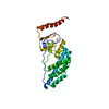
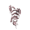
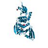
- PDB-5ow4: Crystal structure of a protease-resistant fragment of the Trypano... -
+ Open data

- Basic information
| Entry | Database: PDB / ID: 5ow4 | ||||||
|---|---|---|---|---|---|---|---|
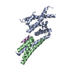
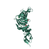
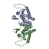
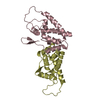
| Title | Crystal structure of a protease-resistant fragment of the Trypanosoma cruzi gamete fusion protein HAP2 ectodomain | ||||||
 Components Components | Uncharacterized protein | ||||||
 Keywords Keywords | MEMBRANE PROTEIN / Gamete fusion protein / class II fold / HAP2 / Membrane fusion | ||||||
| Function / homology | Generative cell specific-1/HAP2 domain / Male gamete fusion factor / HAP2/GCS1 / single fertilization / lipid binding / plasma membrane / Generative cell specific-1/HAP2 domain-containing protein Function and homology information Function and homology information | ||||||
| Biological species |  | ||||||
| Method |  X-RAY DIFFRACTION / SYNCHROTRON / MOLECULAR REPLACEMENT / Resolution: 3.1 Å X-RAY DIFFRACTION / SYNCHROTRON / MOLECULAR REPLACEMENT / Resolution: 3.1 Å | ||||||
 Authors Authors | Fedry, J. / Rey, F.A. / Krey, T. | ||||||
| Funding support |  France, 1items France, 1items
| ||||||
 Citation Citation | Journal: PLoS Biol. / Year: 2018 Title: Evolutionary diversification of the HAP2 membrane insertion motifs to drive gamete fusion across eukaryotes. Authors: Fedry, J. / Forcina, J. / Legrand, P. / Pehau-Arnaudet, G. / Haouz, A. / Johnson, M. / Rey, F.A. / Krey, T. | ||||||
| History |
|
- Structure visualization
| Structure viewer | Molecule: MolmilJmol/JSmol |
|---|
- Downloads & links
-Download
| PDBx/mmCIF format | 5ow4.cif.gz | 68.7 KB | Display | PDBx/mmCIF format |
|---|---|---|---|---|
| PDB format | pdb5ow4.ent.gz | 45.1 KB | Display | PDB format |
| PDBx/mmJSON format | 5ow4.json.gz | Tree view | PDBx/mmJSON format | |
| Others |  Other downloads Other downloads |
-Validation report
| Arichive directory | https://data.pdbj.org/pub/pdb/validation_reports/ow/5ow4ftp://data.pdbj.org/pub/pdb/validation_reports/ow/5ow4 | HTTPS FTP |
|---|
-Related structure data
| Related structure data | 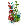 5ow3C  5mf1S S: Starting model for refinement C: citing same article ( |
|---|---|
| Similar structure data |


-Links
 PDBj
PDBj- Assembly
| Deposited unit | 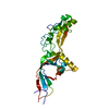
| ||||||||
|---|---|---|---|---|---|---|---|---|---|
| 1 |
| ||||||||
| Unit cell |
|
-Components
| #1: Protein | Mass: 64553.969 Da / Num. of mol.: 1 Source method: isolated from a genetically manipulated source Source: (gene. exp.) Gene: Tc00.1047053509105.4 / Cell line (production host): Schneider 2 / Production host:  |
|---|---|
| #2: Water | ChemComp-HOH /  Mass: 18.015 Da / Num. of mol.: 2 / Source method: isolated from a natural source / Formula: H2O Mass: 18.015 Da / Num. of mol.: 2 / Source method: isolated from a natural source / Formula: H2O |
| Has protein modification | Y |
-Experimental details
-Experiment
| Experiment | Method: X-RAY DIFFRACTION / Number of used crystals: 1 |
|---|
- Sample preparation
| Crystal | Density Matthews: 3.12 Å3/Da / Density % sol: 60.54 % |
|---|---|
| Crystal grow | Temperature: 293 K / Method: vapor diffusion, hanging drop / pH: 9 / Details: 100mM CHES pH9.0 200mM NaCl 10%w/v PEG 8K |
-Data collection
| Diffraction | Mean temperature: 110 K |
|---|---|
| Diffraction source | Source: SYNCHROTRON / Site: SLS  / Beamline: X06SA / Wavelength: 1 Å / Beamline: X06SA / Wavelength: 1 Å |
| Detector | Type: DECTRIS PILATUS3 6M / Detector: PIXEL / Date: Mar 15, 2015 |
| Radiation | Protocol: SINGLE WAVELENGTH / Monochromatic (M) / Laue (L): M / Scattering type: x-ray |
| Radiation wavelength | Wavelength: 1 Å / Relative weight: 1 |
| Reflection | Resolution: 3.1→50 Å / Num. obs: 15453 / % possible obs: 97.7 % / Redundancy: 8.9 % / Biso Wilson estimate: 116.25 Å2 / CC1/2: 0.997 / Rrim(I) all: 0.201 / Net I/σ(I): 11.4 |
| Reflection shell | Resolution: 3.1→3.31 Å / Redundancy: 8.6 % / Mean I/σ(I) obs: 1.3 / Num. unique obs: 2389 / CC1/2: 0.41 / Rrim(I) all: 2.493 / % possible all: 98.2 |
- Processing
| Software |
| ||||||||||||||||||||||||||||||||||||||||||||||||||||||||||||||||||||||||||||||||||||||||||||||||||||||||||||||||||
|---|---|---|---|---|---|---|---|---|---|---|---|---|---|---|---|---|---|---|---|---|---|---|---|---|---|---|---|---|---|---|---|---|---|---|---|---|---|---|---|---|---|---|---|---|---|---|---|---|---|---|---|---|---|---|---|---|---|---|---|---|---|---|---|---|---|---|---|---|---|---|---|---|---|---|---|---|---|---|---|---|---|---|---|---|---|---|---|---|---|---|---|---|---|---|---|---|---|---|---|---|---|---|---|---|---|---|---|---|---|---|---|---|---|---|---|
| Refinement | Method to determine structure: MOLECULAR REPLACEMENT Starting model: 5MF1 Resolution: 3.1→49.13 Å / Cor.coef. Fo:Fc: 0.8853 / Cor.coef. Fo:Fc free: 0.8521 / SU R Cruickshank DPI: 0.346 / Cross valid method: THROUGHOUT / σ(F): 0 / SU R Blow DPI: 0.349 / SU Rfree Blow DPI: 0.277 / SU Rfree Cruickshank DPI: 0.278
| ||||||||||||||||||||||||||||||||||||||||||||||||||||||||||||||||||||||||||||||||||||||||||||||||||||||||||||||||||
| Displacement parameters | Biso mean: 84.33 Å2
| ||||||||||||||||||||||||||||||||||||||||||||||||||||||||||||||||||||||||||||||||||||||||||||||||||||||||||||||||||
| Refine analyze | Luzzati coordinate error obs: 0.659 Å | ||||||||||||||||||||||||||||||||||||||||||||||||||||||||||||||||||||||||||||||||||||||||||||||||||||||||||||||||||
| Refinement step | Cycle: 1 / Resolution: 3.1→49.13 Å
| ||||||||||||||||||||||||||||||||||||||||||||||||||||||||||||||||||||||||||||||||||||||||||||||||||||||||||||||||||
| Refine LS restraints |
| ||||||||||||||||||||||||||||||||||||||||||||||||||||||||||||||||||||||||||||||||||||||||||||||||||||||||||||||||||
| LS refinement shell | Resolution: 3.1→3.31 Å / Total num. of bins used: 8
|
