 Movie
Movie Controller


+ Open data

- Basic information
| Entry | Database: PDB / ID: 2xrh | ||||||
|---|---|---|---|---|---|---|---|
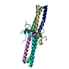
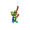
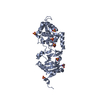
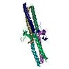
| Title | Crystal structure of the truncated form of HP0721 | ||||||
 Components Components | PROTEIN HP0721 | ||||||
 Keywords Keywords | UNKNOWN FUNCTION | ||||||
| Function / homology | HP0721 helical bundle / Protein of unknown function DUF1104 / DUF1104 superfamily / Protein of unknown function (DUF1104) / Four Helix Bundle (Hemerythrin (Met), subunit A) / Up-down Bundle / Mainly Alpha / NICOTINIC ACID / Zinc ABC transporter substrate-binding protein Function and homology information Function and homology information | ||||||
| Biological species |   HELICOBACTER PYLORI (bacteria) HELICOBACTER PYLORI (bacteria) | ||||||
| Method |  X-RAY DIFFRACTION / SYNCHROTRON / MAD / Resolution: 1.5 Å X-RAY DIFFRACTION / SYNCHROTRON / MAD / Resolution: 1.5 Å | ||||||
 Authors Authors | Cioci, G. / Terradot, L. / Dian, C. / Muller-Dieckmann, C. / Leonard, G. | ||||||
 Citation Citation | Journal: Proteins / Year: 2011 Title: Crystal Structure of Hp0721, a Novel Secreted Protein from Helicobacter Pylori. Authors: Cioci, G. / Terradot, L. / Dian, C. / Mueller-Dieckmann, C. / Leonard, G. | ||||||
| History |
|
- Structure visualization
| Structure viewer | Molecule: MolmilJmol/JSmol |
|---|
- Downloads & links
-Download
| PDBx/mmCIF format | 2xrh.cif.gz | 59.2 KB | Display | PDBx/mmCIF format |
|---|---|---|---|---|
| PDB format | pdb2xrh.ent.gz | 44.2 KB | Display | PDB format |
| PDBx/mmJSON format | 2xrh.json.gz | Tree view | PDBx/mmJSON format | |
| Others |  Other downloads Other downloads |
-Validation report
| Arichive directory | https://data.pdbj.org/pub/pdb/validation_reports/xr/2xrhftp://data.pdbj.org/pub/pdb/validation_reports/xr/2xrh | HTTPS FTP |
|---|
-Related structure data
| Similar structure data |
|---|

-Links
 PDBj
PDBj- Assembly
| Deposited unit | 
| ||||||||||||||||||
|---|---|---|---|---|---|---|---|---|---|---|---|---|---|---|---|---|---|---|---|
| 1 | 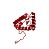
| ||||||||||||||||||
| Unit cell |
| ||||||||||||||||||
| Components on special symmetry positions |
|
-Components
| #1: Protein | Mass: 11512.160 Da / Num. of mol.: 1 / Fragment: RESIDUES 20-112 Source method: isolated from a genetically manipulated source Source: (gene. exp.) HELICOBACTER PYLORI (bacteria) / Strain: 26695 / Plasmid: PET151/DTOPO / Production host: |
|---|---|
| #2: Chemical | ChemComp-NIO /   Mass: 123.109 Da / Num. of mol.: 1 / Source method: obtained synthetically / Formula: C6H5NO2 / Comment: medication*YM Mass: 123.109 Da / Num. of mol.: 1 / Source method: obtained synthetically / Formula: C6H5NO2 / Comment: medication*YM |
| #3: Water | ChemComp-HOH /  Mass: 18.015 Da / Num. of mol.: 112 / Source method: isolated from a natural source / Formula: H2O Mass: 18.015 Da / Num. of mol.: 112 / Source method: isolated from a natural source / Formula: H2O |
| Nonpolymer details | NICOTINIC ACID (NIO): ENDOGENOUS |
-Experimental details
-Experiment
| Experiment | Method: X-RAY DIFFRACTION / Number of used crystals: 1 |
|---|
- Sample preparation
| Crystal | Density Matthews: 2.3 Å3/Da / Density % sol: 46 % / Description: NONE |
|---|---|
| Crystal grow | pH: 7 / Details: 2.9M SODIUM MALONATE, PH 7 |
-Data collection
| Diffraction | Mean temperature: 100 K | |||||||||
|---|---|---|---|---|---|---|---|---|---|---|
| Diffraction source | Source: SYNCHROTRON / Site: ESRF  / Beamline: ID29 / Wavelength: 0.954, 1.907 / Beamline: ID29 / Wavelength: 0.954, 1.907 | |||||||||
| Detector | Type: ADSC CCD / Detector: CCD / Details: MIRRORS | |||||||||
| Radiation | Monochromator: SI111 / Protocol: MAD / Monochromatic (M) / Laue (L): M / Scattering type: x-ray | |||||||||
| Radiation wavelength |
| |||||||||
| Reflection | Resolution: 1.5→41.38 Å / Num. obs: 17909 / % possible obs: 98.6 % / Observed criterion σ(I): 2 / Redundancy: 5.11 % / Biso Wilson estimate: 17.636 Å2 / Rmerge(I) obs: 0.05 / Net I/σ(I): 11.58 | |||||||||
| Reflection shell | Resolution: 1.5→1.58 Å / Redundancy: 4.06 % / Rmerge(I) obs: 0.48 / Mean I/σ(I) obs: 1.6 / % possible all: 93.2 |
- Processing
| Software |
| ||||||||||||||||||||||||||||||||||||||||||||||||||||||||||||||||||||||||||||||||||||||||||||||||||||||||||||||||||||||||||||||||||||||||||||||||||||||||||||||||||||||||||||||||||||||
|---|---|---|---|---|---|---|---|---|---|---|---|---|---|---|---|---|---|---|---|---|---|---|---|---|---|---|---|---|---|---|---|---|---|---|---|---|---|---|---|---|---|---|---|---|---|---|---|---|---|---|---|---|---|---|---|---|---|---|---|---|---|---|---|---|---|---|---|---|---|---|---|---|---|---|---|---|---|---|---|---|---|---|---|---|---|---|---|---|---|---|---|---|---|---|---|---|---|---|---|---|---|---|---|---|---|---|---|---|---|---|---|---|---|---|---|---|---|---|---|---|---|---|---|---|---|---|---|---|---|---|---|---|---|---|---|---|---|---|---|---|---|---|---|---|---|---|---|---|---|---|---|---|---|---|---|---|---|---|---|---|---|---|---|---|---|---|---|---|---|---|---|---|---|---|---|---|---|---|---|---|---|---|---|
| Refinement | Method to determine structure: MAD Starting model: NONE Resolution: 1.5→41.38 Å / Cor.coef. Fo:Fc: 0.961 / Cor.coef. Fo:Fc free: 0.946 / SU B: 2.892 / SU ML: 0.049 / Cross valid method: THROUGHOUT / ESU R: 0.092 / ESU R Free: 0.077 / Stereochemistry target values: MAXIMUM LIKELIHOOD Details: HYDROGENS HAVE BEEN ADDED IN THE RIDING POSITIONS. U VALUES REFINED INDIVIDUALLY
| ||||||||||||||||||||||||||||||||||||||||||||||||||||||||||||||||||||||||||||||||||||||||||||||||||||||||||||||||||||||||||||||||||||||||||||||||||||||||||||||||||||||||||||||||||||||
| Solvent computation | Ion probe radii: 0.8 Å / Shrinkage radii: 0.8 Å / VDW probe radii: 1.2 Å / Solvent model: MASK | ||||||||||||||||||||||||||||||||||||||||||||||||||||||||||||||||||||||||||||||||||||||||||||||||||||||||||||||||||||||||||||||||||||||||||||||||||||||||||||||||||||||||||||||||||||||
| Displacement parameters | Biso mean: 16.761 Å2
| ||||||||||||||||||||||||||||||||||||||||||||||||||||||||||||||||||||||||||||||||||||||||||||||||||||||||||||||||||||||||||||||||||||||||||||||||||||||||||||||||||||||||||||||||||||||
| Refinement step | Cycle: LAST / Resolution: 1.5→41.38 Å
| ||||||||||||||||||||||||||||||||||||||||||||||||||||||||||||||||||||||||||||||||||||||||||||||||||||||||||||||||||||||||||||||||||||||||||||||||||||||||||||||||||||||||||||||||||||||
| Refine LS restraints |
|