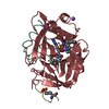
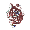
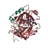
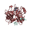
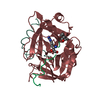
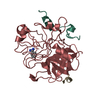
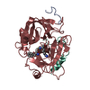
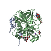
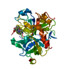
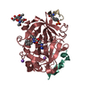
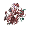
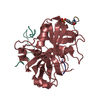
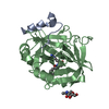
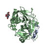
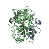
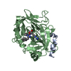
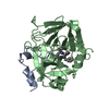
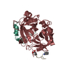
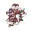
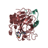
#1: Journal: Protein Expr.Purif. / Year: 1997 Title: Stable Expression and Purification of a Secreted Human Recombinant Prethrombin-2 and its Activation to Thrombin Authors: Russo, G. / Gast, A. / Schlaeger, E.J. / Angiolillo, A. / Pietropaolo, C.
History
Deposition
May 24, 2004
Deposition site: PDBE / Processing site: PDBE
Revision 1.0
Jun 20, 2005
Provider: repository / Type: Initial release
Revision 1.1
Jul 13, 2011
Group: Atomic model / Database references ...Atomic model / Database references / Derived calculations / Non-polymer description / Structure summary / Version format compliance
Revision 1.2
Jun 28, 2017
Group: Data collection / Category: diffrn_source / Item: _diffrn_source.type
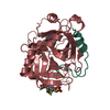
Resolution: 1.54→18.55 Å / Rfactor Rfree error: 0.004 / Isotropic thermal model: RESTRAINED / Cross valid method: THROUGHOUT / σ(F): 0 Details: CHYMOTRYPSIN NUMBERING RATHER THAN SEQUENTIAL SYSTEM IS USED, BASED ON THE TOPOLOGICAL ALIGNMENT WITH THE STRUCTURE OF CHYMOTRYPSIN W.BODE ET AL., 1989, EMBO J.8, 3467-3475. THE STRUCTURE OF ...Details: CHYMOTRYPSIN NUMBERING RATHER THAN SEQUENTIAL SYSTEM IS USED, BASED ON THE TOPOLOGICAL ALIGNMENT WITH THE STRUCTURE OF CHYMOTRYPSIN W.BODE ET AL., 1989, EMBO J.8, 3467-3475. THE STRUCTURE OF 1OYT.PDB WAS TAKEN AS STARTING POINT FOR THE REFINEMENT. APART FROM THE INHIBITOR AND A FEW WATER MOLECULES THE TWO STRUCTURES ARE THE SAME. WATER S339 HAS ELECTRON DENSITY, BUT IS NOT RELIABLE ON GEOMETRIC GROUNDS. OTHER WATER MOLECULES WITH B OVER 50 SHOULD NOT BE TRUSTED. THE INHIBITOR AND GLU 192 HAVE GOOD DENSITY, SO THE SHORT HYDROGEN BOND BETWEEN THEM IS RELIABLE
In the structure databanks used in Yorodumi, some data are registered as the other names, "COVID-19 virus" and "2019-nCoV". Here are the details of the virus and the list of structure data.
Jan 31, 2019. EMDB accession codes are about to change! (news from PDBe EMDB page)
EMDB accession codes are about to change! (news from PDBe EMDB page)
The allocation of 4 digits for EMDB accession codes will soon come to an end. Whilst these codes will remain in use, new EMDB accession codes will include an additional digit and will expand incrementally as the available range of codes is exhausted. The current 4-digit format prefixed with “EMD-” (i.e. EMD-XXXX) will advance to a 5-digit format (i.e. EMD-XXXXX), and so on. It is currently estimated that the 4-digit codes will be depleted around Spring 2019, at which point the 5-digit format will come into force.
The EM Navigator/Yorodumi systems omit the EMD- prefix.
Related info.:Q: What is EMD? / ID/Accession-code notation in Yorodumi/EM Navigator
Yorodumi is a browser for structure data from EMDB, PDB, SASBDB, etc.
This page is also the successor to EM Navigator detail page, and also detail information page/front-end page for Omokage search.
The word "yorodu" (or yorozu) is an old Japanese word meaning "ten thousand". "mi" (miru) is to see.
Related info.:EMDB / PDB / SASBDB / Comparison of 3 databanks / Yorodumi Search / Aug 31, 2016. New EM Navigator & Yorodumi / Yorodumi Papers / Jmol/JSmol / Function and homology information / Changes in new EM Navigator and Yorodumi
 Movie
Movie Controller


 Open data
 Basic information
 Components
 Keywords
 Function and homology information
 HOMO SAPIENS (human)
 HIRUDO MEDICINALIS (medicinal leech)
 X-RAY DIFFRACTION /
  Authors
 Citation
 Structure visualization
 Downloads & links
 Other downloads


 PDBj
PDBj


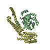
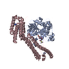
 Assembly

 CRICETULUS GRISEUS (Chinese hamster) / References: UniProt: P00734, thrombin


 Mass: 448.471 Da / Num. of mol.: 1 / Source method: obtained synthetically / Formula: C24H24N4O5
 Mass: 22.990 Da / Num. of mol.: 1 / Source method: obtained synthetically / Formula: Na
 Mass: 40.078 Da / Num. of mol.: 1 / Source method: obtained synthetically / Formula: Ca
 Sample preparation
 Processing