 Movie
Movie Controller

[English] 日本語
 Yorodumi
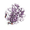
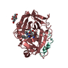
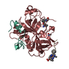
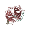
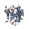
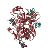
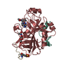
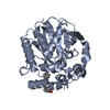
- PDB-1hah: THE ISOMORPHOUS STRUCTURES OF PRETHROMBIN2, HIRUGEN-AND PPACK-THR... -
+ Open data

- Basic information
| Entry | Database: PDB / ID: 1hah | ||||||
|---|---|---|---|---|---|---|---|
| Title | THE ISOMORPHOUS STRUCTURES OF PRETHROMBIN2, HIRUGEN-AND PPACK-THROMBIN: CHANGES ACCOMPANYING ACTIVATION AND EXOSITE BINDING TO THROMBIN | ||||||
 Components Components |
| ||||||
 Keywords Keywords | COMPLEX(SERINE PROTEINASE/INHIBITOR) / COMPLEX(SERINE PROTEINASE-INHIBITOR) / COMPLEX(SERINE PROTEINASE-INHIBITOR) complex | ||||||
| Function / homology |  Function and homology information Function and homology information: / thrombospondin receptor activity / thrombin / thrombin-activated receptor signaling pathway / Defective factor XII causes hereditary angioedema / negative regulation of astrocyte differentiation / regulation of blood coagulation / neutrophil-mediated killing of gram-negative bacterium / positive regulation of phospholipase C-activating G protein-coupled receptor signaling pathway / Defective F8 cleavage by thrombin ...: / thrombospondin receptor activity / thrombin / thrombin-activated receptor signaling pathway / Defective factor XII causes hereditary angioedema / negative regulation of astrocyte differentiation / regulation of blood coagulation / neutrophil-mediated killing of gram-negative bacterium / positive regulation of phospholipase C-activating G protein-coupled receptor signaling pathway / Defective F8 cleavage by thrombin / ligand-gated ion channel signaling pathway / Platelet Aggregation (Plug Formation) / positive regulation of collagen biosynthetic process / negative regulation of platelet activation / negative regulation of blood coagulation / negative regulation of fibrinolysis / blood coagulation, fibrin clot formation / positive regulation of blood coagulation / Transport of gamma-carboxylated protein precursors from the endoplasmic reticulum to the Golgi apparatus / : / Gamma-carboxylation of protein precursors / Removal of aminoterminal propeptides from gamma-carboxylated proteins / regulation of cytosolic calcium ion concentration / fibrinolysis / : / negative regulation of proteolysis / negative regulation of cytokine production involved in inflammatory response / Regulation of Complement cascade / positive regulation of release of sequestered calcium ion into cytosol / acute-phase response / Cell surface interactions at the vascular wall / Peptide ligand-binding receptors / growth factor activity / positive regulation of receptor signaling pathway via JAK-STAT / serine-type endopeptidase inhibitor activity / lipopolysaccharide binding / platelet activation / positive regulation of protein localization to nucleus / response to wounding / Golgi lumen / Regulation of Insulin-like Growth Factor (IGF) transport and uptake by Insulin-like Growth Factor Binding Proteins (IGFBPs) / positive regulation of reactive oxygen species metabolic process / blood coagulation / positive regulation of insulin secretion / regulation of cell shape / antimicrobial humoral immune response mediated by antimicrobial peptide / heparin binding / Thrombin signalling through proteinase activated receptors (PARs) / positive regulation of cell growth / blood microparticle / G alpha (q) signalling events / positive regulation of phosphatidylinositol 3-kinase/protein kinase B signal transduction / cell surface receptor signaling pathway / endoplasmic reticulum lumen / receptor ligand activity / serine-type endopeptidase activity / signaling receptor binding / calcium ion binding / positive regulation of cell population proliferation / proteolysis / : / extracellular exosome / extracellular region / plasma membrane Similarity search - Function | ||||||
| Biological species |  Homo sapiens (human) Homo sapiens (human) Hirudo medicinalis (medicinal leech) Hirudo medicinalis (medicinal leech) | ||||||
| Method |  X-RAY DIFFRACTION / Resolution: 2.3 Å X-RAY DIFFRACTION / Resolution: 2.3 Å | ||||||
 Authors Authors | Tulinsky, A. / Vijayalakshmi, J. | ||||||
 Citation Citation | Journal: Protein Sci. / Year: 1994 Title: The isomorphous structures of prethrombin2, hirugen-, and PPACK-thrombin: changes accompanying activation and exosite binding to thrombin. Authors: Vijayalakshmi, J. / Padmanabhan, K.P. / Mann, K.G. / Tulinsky, A. #1: Journal: J.Mol.Biol. / Year: 1991Title: Refined Structure of the Hirudin-Thrombin Complex Authors: Rydel, T.J. / Tulinsky, A. / Bode, W. / Huber, R. #2: Journal: J.Mol.Biol. / Year: 1991Title: Structure of the Hirugen and Hirulog 1 Complexes of Alpha-Thrombin Authors: Skrzypczak-Jankun, E. / Carperos, V.E. / Ravichandran, K.G. / Tulinsky, A. / Westbrook, M. / Maraganore, J.M. | ||||||
| History |
| ||||||
| Remark 700 | SHEET THE SHEET PRESENTED AS *B1* ON SHEET RECORDS BELOW IS ACTUALLY A SIX-STRANDED BETA-BARREL. ...SHEET THE SHEET PRESENTED AS *B1* ON SHEET RECORDS BELOW IS ACTUALLY A SIX-STRANDED BETA-BARREL. THIS IS REPRESENTED BY A SEVEN-STRANDED SHEET IN WHICH THE FIRST AND LAST STRANDS ARE IDENTICAL. THE SHEET PRESENTED AS *B2* ON SHEET RECORDS BELOW IS ACTUALLY A SEVEN-STRANDED BETA-BARREL. THIS IS REPRESENTED BY AN EIGHT-STRANDED SHEET IN WHICH THE FIRST AND LAST STRANDS ARE IDENTICAL. |
- Structure visualization
| Structure viewer | Molecule: MolmilJmol/JSmol |
|---|
- Downloads & links
-Download
| PDBx/mmCIF format | 1hah.cif.gz | 81.5 KB | Display | PDBx/mmCIF format |
|---|---|---|---|---|
| PDB format | pdb1hah.ent.gz | 59.8 KB | Display | PDB format |
| PDBx/mmJSON format | 1hah.json.gz | Tree view | PDBx/mmJSON format | |
| Others |  Other downloads Other downloads |
-Validation report
| Arichive directory | https://data.pdbj.org/pub/pdb/validation_reports/ha/1hahftp://data.pdbj.org/pub/pdb/validation_reports/ha/1hah | HTTPS FTP |
|---|
-Related structure data

-Links
 PDBj
PDBj


- Assembly
| Deposited unit | 
| |||||||||
|---|---|---|---|---|---|---|---|---|---|---|
| 1 |
| |||||||||
| Unit cell |
| |||||||||
| Atom site foot note | 1: CIS PROLINE - PRO H 37 2: RESIDUE TYR I 63 IN THE HIRUGEN MOLECULE IS BONDED TO SULFATO- TYROSINE. | |||||||||
| Components on special symmetry positions |
|
-Components
| #1: Protein/peptide | Mass: 4096.534 Da / Num. of mol.: 1 Source method: isolated from a genetically manipulated source Source: (gene. exp.) Homo sapiens (human) / References: UniProt: P00734, thrombin |
|---|---|
| #2: Protein | Mass: 29780.219 Da / Num. of mol.: 1 Source method: isolated from a genetically manipulated source Source: (gene. exp.) Homo sapiens (human) / References: UniProt: P00734, thrombin |
| #3: Protein/peptide | Mass: 1363.399 Da / Num. of mol.: 1 Source method: isolated from a genetically manipulated source Source: (gene. exp.) Hirudo medicinalis (medicinal leech) / References: UniProt: P28507*PLUS |
| #4: Sugar | ChemComp-NAG /   Type: D-saccharide, beta linking / Mass: 221.208 Da / Num. of mol.: 1 Type: D-saccharide, beta linking / Mass: 221.208 Da / Num. of mol.: 1Source method: isolated from a genetically manipulated source Formula: C8H15NO6 |
| #5: Water | ChemComp-HOH /  Mass: 18.015 Da / Num. of mol.: 204 / Source method: isolated from a natural source / Formula: H2O Mass: 18.015 Da / Num. of mol.: 204 / Source method: isolated from a natural source / Formula: H2O |
| Compound details | THROMBIN IS CLEAVED BETWEEN RESIDUES 15 AND 16. CHAIN INDICATOR *L* IS USED FOR RESIDUES 1H - 15 ...THROMBIN IS CLEAVED BETWEEN RESIDUES 15 AND 16. CHAIN INDICATOR *L* IS USED FOR RESIDUES 1H - 15 AND CHAIN INDICATOR *H* IS USED FOR RESIDUES 16 - 247. CHAIN INDICATOR *I* IS USED FOR N-ACETYLHIRU |
| Has protein modification | Y |
| Sequence details | PEPTIDE ASP I 55 - LEU I 64 AT THE EXOSITE OF THROMBIN IS HIRUGEN MOLECULE, WHICH IS PART OF HIRUDIN 53 - 65. |
-Experimental details
-Experiment
| Experiment | Method: X-RAY DIFFRACTION |
|---|
- Sample preparation
| Crystal | Density Matthews: 2.66 Å3/Da / Density % sol: 53.79 % | ||||||||||||||||||||||||||||||||||||||||||||||||||||||
|---|---|---|---|---|---|---|---|---|---|---|---|---|---|---|---|---|---|---|---|---|---|---|---|---|---|---|---|---|---|---|---|---|---|---|---|---|---|---|---|---|---|---|---|---|---|---|---|---|---|---|---|---|---|---|---|
| Crystal grow | *PLUS pH: 7.3 / Method: microdialysis | ||||||||||||||||||||||||||||||||||||||||||||||||||||||
| Components of the solutions | *PLUS
|
-Data collection
| Radiation | Scattering type: x-ray |
|---|---|
| Radiation wavelength | Relative weight: 1 |
| Reflection | *PLUS Highest resolution: 2.3 Å / Num. obs: 15157 / % possible obs: 83 % / Rmerge(I) obs: 0.068 |
- Processing
| Software |
| ||||||||||||||||||||||||||||||||||||||||||||||||||||||||||||||||||||||||||||||||
|---|---|---|---|---|---|---|---|---|---|---|---|---|---|---|---|---|---|---|---|---|---|---|---|---|---|---|---|---|---|---|---|---|---|---|---|---|---|---|---|---|---|---|---|---|---|---|---|---|---|---|---|---|---|---|---|---|---|---|---|---|---|---|---|---|---|---|---|---|---|---|---|---|---|---|---|---|---|---|---|---|---|
| Refinement | Resolution: 2.3→7 Å / σ(F): 2 /
| ||||||||||||||||||||||||||||||||||||||||||||||||||||||||||||||||||||||||||||||||
| Refinement step | Cycle: LAST / Resolution: 2.3→7 Å
| ||||||||||||||||||||||||||||||||||||||||||||||||||||||||||||||||||||||||||||||||
| Refine LS restraints |
| ||||||||||||||||||||||||||||||||||||||||||||||||||||||||||||||||||||||||||||||||
| Software | *PLUS Name: X-PLOR/PROLSQ / Classification: refinement | ||||||||||||||||||||||||||||||||||||||||||||||||||||||||||||||||||||||||||||||||
| Refinement | *PLUS Rfactor obs: 0.114 | ||||||||||||||||||||||||||||||||||||||||||||||||||||||||||||||||||||||||||||||||
| Solvent computation | *PLUS | ||||||||||||||||||||||||||||||||||||||||||||||||||||||||||||||||||||||||||||||||
| Displacement parameters | *PLUS Biso mean: 27.7 Å2 | ||||||||||||||||||||||||||||||||||||||||||||||||||||||||||||||||||||||||||||||||
| Refine LS restraints | *PLUS
|
