 Movie
Movie Controller

[English] 日本語
 Yorodumi
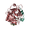
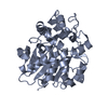
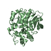
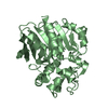
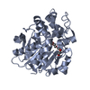
- PDB-4uhb: Laboratory evolved variant R-C1 of potato epoxide hydrolase StEH1 -
+ Open data

- Basic information
| Entry | Database: PDB / ID: 4uhb | ||||||
|---|---|---|---|---|---|---|---|
| Title | Laboratory evolved variant R-C1 of potato epoxide hydrolase StEH1 | ||||||
 Components Components | EPOXIDE HYDROLASE | ||||||
 Keywords Keywords | HYDROLASE / DIRECTED EVOLUTION | ||||||
| Function / homology |  Function and homology information Function and homology information | ||||||
| Biological species |  | ||||||
| Method |  X-RAY DIFFRACTION / SYNCHROTRON / MOLECULAR REPLACEMENT / Resolution: 1.8 Å X-RAY DIFFRACTION / SYNCHROTRON / MOLECULAR REPLACEMENT / Resolution: 1.8 Å | ||||||
 Authors Authors | Nilsson, M.T.I. / Carlsson, A.J. / Dobritzsch, D. / Widersten, M. | ||||||
 Citation Citation | Journal: Chembiochem / Year: 2016 Title: Laboratory Evolved Enzymes Provide Snapshots of the Development of Enantioconvergence in Enzyme-Catalyzed Epoxide Hydrolysis. Authors: Janfalk Carlsson, A. / Bauer, P. / Dobritzsch, D. / Nilsson, M. / Kamerlin, S.C. / Widersten, M. | ||||||
| History |
|
- Structure visualization
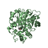
| Structure viewer | Molecule: MolmilJmol/JSmol |
|---|
- Downloads & links
-Download
| PDBx/mmCIF format | 4uhb.cif.gz | 270 KB | Display | PDBx/mmCIF format |
|---|---|---|---|---|
| PDB format | pdb4uhb.ent.gz | 221.8 KB | Display | PDB format |
| PDBx/mmJSON format | 4uhb.json.gz | Tree view | PDBx/mmJSON format | |
| Others |  Other downloads Other downloads |
-Validation report
| Arichive directory | https://data.pdbj.org/pub/pdb/validation_reports/uh/4uhbftp://data.pdbj.org/pub/pdb/validation_reports/uh/4uhb | HTTPS FTP |
|---|
-Related structure data
| Related structure data |  4ufoC 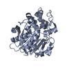 4ufpC 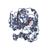 2cjpS S: Starting model for refinement C: citing same article ( |
|---|---|
| Similar structure data |


-Links
 PDBj
PDBj


- Assembly
| Deposited unit | 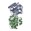
| ||||||||||||||||||
|---|---|---|---|---|---|---|---|---|---|---|---|---|---|---|---|---|---|---|---|
| 1 | 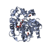
| ||||||||||||||||||
| 2 | 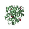
| ||||||||||||||||||
| Unit cell |
| ||||||||||||||||||
| Noncrystallographic symmetry (NCS) | NCS domain:
NCS domain segments: Component-ID: _ / Ens-ID: 1 / Beg auth comp-ID: LYS / Beg label comp-ID: LYS / End auth comp-ID: LYS / End label comp-ID: LYS / Refine code: _ / Auth seq-ID: 3 - 320 / Label seq-ID: 3 - 320
NCS oper: (Code: given Matrix: (0.7088, 0.6543, -0.2637), Vector: |
-Components
| #1: Protein | Mass: 37171.562 Da / Num. of mol.: 2 / Mutation: YES Source method: isolated from a genetically manipulated source Source: (gene. exp.)  #2: Chemical | ChemComp-GOL /   Mass: 92.094 Da / Num. of mol.: 9 / Source method: obtained synthetically / Formula: C3H8O3 Mass: 92.094 Da / Num. of mol.: 9 / Source method: obtained synthetically / Formula: C3H8O3#3: Chemical |   Mass: 62.068 Da / Num. of mol.: 2 / Source method: obtained synthetically / Formula: C2H6O2 Mass: 62.068 Da / Num. of mol.: 2 / Source method: obtained synthetically / Formula: C2H6O2#4: Water | ChemComp-HOH / |  Mass: 18.015 Da / Num. of mol.: 591 / Source method: isolated from a natural source / Formula: H2O Mass: 18.015 Da / Num. of mol.: 591 / Source method: isolated from a natural source / Formula: H2O |
|---|
-Experimental details
-Experiment
| Experiment | Method: X-RAY DIFFRACTION / Number of used crystals: 1 |
|---|
- Sample preparation
| Crystal | Density Matthews: 2.26 Å3/Da / Density % sol: 46 % / Description: NONE |
|---|---|
| Crystal grow | Details: PROTEIN IN 30 MM TRIS-HCL, PH 7.4 MIXED 1:1 WITH 0.1 M HEPES PH 7.5, 20 % PEG 10,000. THEN SOAKED IN25% (V/V) GLYCEROL, 75 MM HEPES, PH7.68, PEG 10,000 22.5 % (W/V) |
-Data collection
| Diffraction | Mean temperature: 100 K |
|---|---|
| Diffraction source | Source: SYNCHROTRON / Site: ESRF  / Beamline: BM30A / Wavelength: 0.979736 / Beamline: BM30A / Wavelength: 0.979736 |
| Detector | Type: ADSC CCD / Detector: CCD / Date: Jun 28, 2013 |
| Radiation | Protocol: SINGLE WAVELENGTH / Monochromatic (M) / Laue (L): M / Scattering type: x-ray |
| Radiation wavelength | Wavelength: 0.979736 Å / Relative weight: 1 |
| Reflection | Resolution: 1.8→32.8 Å / Num. obs: 61576 / % possible obs: 99.8 % / Observed criterion σ(I): 2 / Redundancy: 6.8 % / Rmerge(I) obs: 0.17 / Net I/σ(I): 6.6 |
| Reflection shell | Resolution: 1.8→1.9 Å / Redundancy: 4.7 % / Rmerge(I) obs: 0.83 / Mean I/σ(I) obs: 2 / % possible all: 99.9 |
- Processing
| Software |
| ||||||||||||||||||||||||||||||||||||||||||||||||||||||||||||||||||||||||||||||||||||||||||||||||||||||||||||||||||||||||||||||||||||||||||||||||||||||||||||||||||||||||||||||||||||||
|---|---|---|---|---|---|---|---|---|---|---|---|---|---|---|---|---|---|---|---|---|---|---|---|---|---|---|---|---|---|---|---|---|---|---|---|---|---|---|---|---|---|---|---|---|---|---|---|---|---|---|---|---|---|---|---|---|---|---|---|---|---|---|---|---|---|---|---|---|---|---|---|---|---|---|---|---|---|---|---|---|---|---|---|---|---|---|---|---|---|---|---|---|---|---|---|---|---|---|---|---|---|---|---|---|---|---|---|---|---|---|---|---|---|---|---|---|---|---|---|---|---|---|---|---|---|---|---|---|---|---|---|---|---|---|---|---|---|---|---|---|---|---|---|---|---|---|---|---|---|---|---|---|---|---|---|---|---|---|---|---|---|---|---|---|---|---|---|---|---|---|---|---|---|---|---|---|---|---|---|---|---|---|---|
| Refinement | Method to determine structure: MOLECULAR REPLACEMENT Starting model: PDB ENTRY 2CJP Resolution: 1.8→32.83 Å / Cor.coef. Fo:Fc: 0.937 / Cor.coef. Fo:Fc free: 0.932 / SU B: 5.803 / SU ML: 0.087 / Cross valid method: THROUGHOUT / ESU R: 0.151 / ESU R Free: 0.124 / Stereochemistry target values: MAXIMUM LIKELIHOOD Details: HYDROGENS HAVE BEEN ADDED IN THE RIDING POSITIONS. HYDROGENS HAVE BEEN ADDED IN THE RIDING POSITIONS U VALUES RESIDUAL ONLY
| ||||||||||||||||||||||||||||||||||||||||||||||||||||||||||||||||||||||||||||||||||||||||||||||||||||||||||||||||||||||||||||||||||||||||||||||||||||||||||||||||||||||||||||||||||||||
| Solvent computation | Ion probe radii: 0.8 Å / Shrinkage radii: 0.8 Å / VDW probe radii: 1.2 Å / Solvent model: MASK | ||||||||||||||||||||||||||||||||||||||||||||||||||||||||||||||||||||||||||||||||||||||||||||||||||||||||||||||||||||||||||||||||||||||||||||||||||||||||||||||||||||||||||||||||||||||
| Displacement parameters | Biso mean: 12.172 Å2
| ||||||||||||||||||||||||||||||||||||||||||||||||||||||||||||||||||||||||||||||||||||||||||||||||||||||||||||||||||||||||||||||||||||||||||||||||||||||||||||||||||||||||||||||||||||||
| Refinement step | Cycle: LAST / Resolution: 1.8→32.83 Å
| ||||||||||||||||||||||||||||||||||||||||||||||||||||||||||||||||||||||||||||||||||||||||||||||||||||||||||||||||||||||||||||||||||||||||||||||||||||||||||||||||||||||||||||||||||||||
| Refine LS restraints |
|