 Movie
Movie Controller

[English] 日本語
 Yorodumi
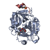
- PDB-3cxu: Structure of a Y149F mutant of epoxide hydrolase from Solanum tub... -
+ Open data

- Basic information
| Entry | Database: PDB / ID: 3cxu | ||||||
|---|---|---|---|---|---|---|---|
| Title | Structure of a Y149F mutant of epoxide hydrolase from Solanum tuberosum | ||||||
 Components Components | Epoxide hydrolase | ||||||
 Keywords Keywords | HYDROLASE / EPOXIDE HYDROLASE / ALPHA/BETA HYDROLASE FOLD / Y149F / PROTON WIRE | ||||||
| Function / homology |  Function and homology information Function and homology information | ||||||
| Biological species |  | ||||||
| Method |  X-RAY DIFFRACTION / SYNCHROTRON / Resolution: 2 Å X-RAY DIFFRACTION / SYNCHROTRON / Resolution: 2 Å | ||||||
 Authors Authors | Naworyta, A. / Mowbray, S.L. / Widersten, M. / Thomaeus, A. | ||||||
 Citation Citation | Journal: Protein Sci. / Year: 2008 Title: Removal of distal protein-water hydrogen bonds in a plant epoxide hydrolase increases catalytic turnover but decreases thermostability Authors: Thomaeus, A. / Naworyta, A. / Mowbray, S.L. / Widersten, M. #1: Journal: Protein Sci. / Year: 2006Title: X-ray structure of potato epoxide hydrolase sheds light on substrate specificity in plant enzymes Authors: Mowbray, S.L. / Elfstrom, L.T. / Ahlgren, K.M. / Andersson, C.E. / Widersten, M. | ||||||
| History |
|
- Structure visualization
| Structure viewer | Molecule: MolmilJmol/JSmol |
|---|
- Downloads & links
-Download
| PDBx/mmCIF format | 3cxu.cif.gz | 152.4 KB | Display | PDBx/mmCIF format |
|---|---|---|---|---|
| PDB format | pdb3cxu.ent.gz | 117.7 KB | Display | PDB format |
| PDBx/mmJSON format | 3cxu.json.gz | Tree view | PDBx/mmJSON format | |
| Others |  Other downloads Other downloads |
-Validation report
| Arichive directory | https://data.pdbj.org/pub/pdb/validation_reports/cx/3cxuftp://data.pdbj.org/pub/pdb/validation_reports/cx/3cxu | HTTPS FTP |
|---|
-Related structure data
| Related structure data | 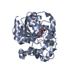 2cjpS S: Starting model for refinement |
|---|---|
| Similar structure data |


-Links
 PDBj
PDBj


- Assembly
| Deposited unit | 
| ||||||||
|---|---|---|---|---|---|---|---|---|---|
| 1 | 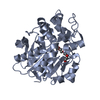
| ||||||||
| 2 | 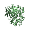
| ||||||||
| Unit cell |
|
-Components
| #1: Protein | Mass: 37139.551 Da / Num. of mol.: 2 / Mutation: Y149F Source method: isolated from a genetically manipulated source Source: (gene. exp.)  #2: Chemical | ChemComp-PG4 / |   Mass: 194.226 Da / Num. of mol.: 1 / Source method: obtained synthetically / Formula: C8H18O5 / Comment: precipitant*YM Mass: 194.226 Da / Num. of mol.: 1 / Source method: obtained synthetically / Formula: C8H18O5 / Comment: precipitant*YM#3: Water | ChemComp-HOH / |  Mass: 18.015 Da / Num. of mol.: 654 / Source method: isolated from a natural source / Formula: H2O Mass: 18.015 Da / Num. of mol.: 654 / Source method: isolated from a natural source / Formula: H2O |
|---|
-Experimental details
-Experiment
| Experiment | Method: X-RAY DIFFRACTION / Number of used crystals: 1 |
|---|
- Sample preparation
| Crystal | Density Matthews: 2.29 Å3/Da / Density % sol: 46.28 % |
|---|---|
| Crystal grow | Temperature: 293 K / Method: vapor diffusion, hanging drop / pH: 7.5 Details: 25% PEG 10000, 0.1M HEPES, pH 7.5, VAPOR DIFFUSION, HANGING DROP, temperature 293K |
-Data collection
| Diffraction | Mean temperature: 100 K |
|---|---|
| Diffraction source | Source: SYNCHROTRON / Site: ESRF  / Beamline: ID23-1 / Wavelength: 1.0723 Å / Beamline: ID23-1 / Wavelength: 1.0723 Å |
| Detector | Type: ADSC QUANTUM 315 / Detector: CCD / Date: Mar 6, 2007 / Details: Single Silicon (111) monochromator |
| Radiation | Monochromator: Single Silicon (111) monochromator / Protocol: SINGLE WAVELENGTH / Monochromatic (M) / Laue (L): M / Scattering type: x-ray |
| Radiation wavelength | Wavelength: 1.0723 Å / Relative weight: 1 |
| Reflection | Resolution: 2→20 Å / Num. obs: 45424 / % possible obs: 99.6 % / Observed criterion σ(I): 0 / Redundancy: 4 % / Biso Wilson estimate: 15.3 Å2 / Rmerge(I) obs: 0.097 / Net I/σ(I): 11.1 |
| Reflection shell | Resolution: 2→2.1 Å / Redundancy: 4.2 % / Rmerge(I) obs: 0.156 / Mean I/σ(I) obs: 8 / Num. unique all: 27087 / % possible all: 99.5 |
- Processing
| Software |
| |||||||||||||||||||||||||||||||||||||||||||||||||||||||||||||||||||||||||||||||||||||||||||||||||||||||||||||||||||||||||||||||||||||||||||||||||||
|---|---|---|---|---|---|---|---|---|---|---|---|---|---|---|---|---|---|---|---|---|---|---|---|---|---|---|---|---|---|---|---|---|---|---|---|---|---|---|---|---|---|---|---|---|---|---|---|---|---|---|---|---|---|---|---|---|---|---|---|---|---|---|---|---|---|---|---|---|---|---|---|---|---|---|---|---|---|---|---|---|---|---|---|---|---|---|---|---|---|---|---|---|---|---|---|---|---|---|---|---|---|---|---|---|---|---|---|---|---|---|---|---|---|---|---|---|---|---|---|---|---|---|---|---|---|---|---|---|---|---|---|---|---|---|---|---|---|---|---|---|---|---|---|---|---|---|---|---|
| Refinement | Starting model: PDB entry 2cjp Resolution: 2→19.976 Å / Cor.coef. Fo:Fc: 0.949 / Cor.coef. Fo:Fc free: 0.926 / WRfactor Rfree: 0.226 / WRfactor Rwork: 0.187 / SU B: 3.206 / SU ML: 0.092 / Isotropic thermal model: isotropic / Cross valid method: THROUGHOUT / σ(F): 0 / ESU R: 0.185 / ESU R Free: 0.152 / Stereochemistry target values: Engh & Huber
| |||||||||||||||||||||||||||||||||||||||||||||||||||||||||||||||||||||||||||||||||||||||||||||||||||||||||||||||||||||||||||||||||||||||||||||||||||
| Solvent computation | Ion probe radii: 0.8 Å / Shrinkage radii: 0.8 Å / VDW probe radii: 1.4 Å / Solvent model: MASK BULK SOLVENT | |||||||||||||||||||||||||||||||||||||||||||||||||||||||||||||||||||||||||||||||||||||||||||||||||||||||||||||||||||||||||||||||||||||||||||||||||||
| Displacement parameters | Biso mean: 14.65 Å2
| |||||||||||||||||||||||||||||||||||||||||||||||||||||||||||||||||||||||||||||||||||||||||||||||||||||||||||||||||||||||||||||||||||||||||||||||||||
| Refine analyze |
| |||||||||||||||||||||||||||||||||||||||||||||||||||||||||||||||||||||||||||||||||||||||||||||||||||||||||||||||||||||||||||||||||||||||||||||||||||
| Refinement step | Cycle: LAST / Resolution: 2→19.976 Å
| |||||||||||||||||||||||||||||||||||||||||||||||||||||||||||||||||||||||||||||||||||||||||||||||||||||||||||||||||||||||||||||||||||||||||||||||||||
| Refine LS restraints |
| |||||||||||||||||||||||||||||||||||||||||||||||||||||||||||||||||||||||||||||||||||||||||||||||||||||||||||||||||||||||||||||||||||||||||||||||||||
| LS refinement shell | Refine-ID: X-RAY DIFFRACTION / Total num. of bins used: 20
|
