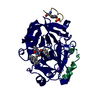
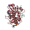
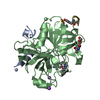
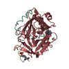
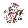
[English] 日本語
Yorodumi
- PDB-2feq: orally active thrombin inhibitors -

+
Open data


ID or keywords:

Loading...

-
Basic information

Entry
Database: PDB / ID: 2feq
Titleorally active thrombin inhibitors
Components
  • Decapeptide Hirudin Analogue
  • Thrombin heavy chain
  • Thrombin light chain
KeywordsHYDROLASE/HYDROLASE INHIBITOR / thrombin inhibitor / hydrolase-hydrolase inhibitor COMPLEX
Function / homology
Function and homology information


positive regulation of lipid kinase activity / positive regulation of phospholipase C-activating G protein-coupled receptor signaling pathway / cytolysis by host of symbiont cells / thrombospondin receptor activity / Defective factor XII causes hereditary angioedema / thrombin / neutrophil-mediated killing of gram-negative bacterium / regulation of blood coagulation / ligand-gated ion channel signaling pathway / Defective F8 cleavage by thrombin ...positive regulation of lipid kinase activity / positive regulation of phospholipase C-activating G protein-coupled receptor signaling pathway / cytolysis by host of symbiont cells / thrombospondin receptor activity / Defective factor XII causes hereditary angioedema / thrombin / neutrophil-mediated killing of gram-negative bacterium / regulation of blood coagulation / ligand-gated ion channel signaling pathway / Defective F8 cleavage by thrombin / Platelet Aggregation (Plug Formation) / negative regulation of platelet activation / negative regulation of astrocyte differentiation / positive regulation of collagen biosynthetic process / negative regulation of cytokine production involved in inflammatory response / positive regulation of blood coagulation / negative regulation of fibrinolysis / Gamma-carboxylation of protein precursors / Transport of gamma-carboxylated protein precursors from the endoplasmic reticulum to the Golgi apparatus / Common Pathway of Fibrin Clot Formation / Removal of aminoterminal propeptides from gamma-carboxylated proteins / fibrinolysis / regulation of cytosolic calcium ion concentration / Intrinsic Pathway of Fibrin Clot Formation / Peptide ligand-binding receptors / positive regulation of release of sequestered calcium ion into cytosol / Regulation of Complement cascade / acute-phase response / Cell surface interactions at the vascular wall / lipopolysaccharide binding / negative regulation of proteolysis / positive regulation of receptor signaling pathway via JAK-STAT / growth factor activity / positive regulation of insulin secretion / platelet activation / response to wounding / Golgi lumen / positive regulation of protein localization to nucleus / Regulation of Insulin-like Growth Factor (IGF) transport and uptake by Insulin-like Growth Factor Binding Proteins (IGFBPs) / positive regulation of reactive oxygen species metabolic process / blood coagulation / antimicrobial humoral immune response mediated by antimicrobial peptide / Thrombin signalling through proteinase activated receptors (PARs) / heparin binding / regulation of cell shape / positive regulation of cell growth / G alpha (q) signalling events / collagen-containing extracellular matrix / blood microparticle / cell surface receptor signaling pathway / positive regulation of phosphatidylinositol 3-kinase/protein kinase B signal transduction / positive regulation of protein phosphorylation / G protein-coupled receptor signaling pathway / endoplasmic reticulum lumen / signaling receptor binding / serine-type endopeptidase activity / calcium ion binding / positive regulation of cell population proliferation / proteolysis / extracellular space / extracellular exosome / extracellular region / plasma membrane
Similarity search - Function
Prothrombin/thrombin / Thrombin light chain / Thrombin light chain domain superfamily / Thrombin light chain / Kringle domain / Kringle / Kringle, conserved site / Kringle superfamily / Kringle domain signature. / Kringle domain profile. ...Prothrombin/thrombin / Thrombin light chain / Thrombin light chain domain superfamily / Thrombin light chain / Kringle domain / Kringle / Kringle, conserved site / Kringle superfamily / Kringle domain signature. / Kringle domain profile. / Kringle domain / Vitamin K-dependent carboxylation/gamma-carboxyglutamic (GLA) domain / Gamma-carboxyglutamic acid-rich (GLA) domain / Gamma-carboxyglutamic acid-rich (GLA) domain superfamily / Vitamin K-dependent carboxylation domain. / Gla domain profile. / Domain containing Gla (gamma-carboxyglutamate) residues. / Kringle-like fold / Serine proteases, trypsin family, histidine active site / Serine proteases, trypsin family, serine active site / Peptidase S1A, chymotrypsin family / Serine proteases, trypsin family, histidine active site. / Serine proteases, trypsin domain profile. / Serine proteases, trypsin family, serine active site. / Trypsin-like serine protease / Serine proteases, trypsin domain / Trypsin / Trypsin-like serine proteases / Thrombin, subunit H / Peptidase S1, PA clan, chymotrypsin-like fold / Peptidase S1, PA clan / Beta Barrel / Mainly Beta
Similarity search - Domain/homology
HIRUDIN ANALOGUE / N-(carboxymethyl)-3-cyclohexyl-D-alanyl-N-({4-[(E)-amino(imino)methyl]-1,3-thiazol-2-yl}methyl)-L-prolinamide / Chem-34P / Prothrombin
Similarity search - Component
Biological speciesHomo sapiens (human)
Hirudo medicinalis (medicinal leech)
Synthetic (others)
MethodX-RAY DIFFRACTION / MOLECULAR REPLACEMENT / Resolution: 2.44 Å
AuthorsMack, H. / Baucke, D. / Hornberger, W. / Lange, U.E.W. / Hoeffken, H.W.
CitationJournal: Bioorg.Med.Chem.Lett. / Year: 2006
Title: Orally active thrombin inhibitors. Part 1: optimization of the P1-moiety
Authors: Mack, H. / Baucke, D. / Hornberger, W. / Lange, U.E.W. / Seitz, W. / Hoeffken, H.W.
History
DepositionDec 16, 2005Deposition site: RCSB / Processing site: RCSB
Revision 1.0Aug 8, 2006Provider: repository / Type: Initial release
Revision 1.1May 1, 2008Group: Version format compliance
Revision 1.2Jul 13, 2011Group: Atomic model / Database references ...Atomic model / Database references / Derived calculations / Non-polymer description / Structure summary / Version format compliance
Revision 1.3Dec 12, 2012Group: Other
Revision 1.4Oct 18, 2017Group: Refinement description / Category: software / Item: _software.name
Revision 1.5Apr 4, 2018Group: Data collection / Category: diffrn_source / Item: _diffrn_source.type

-
Structure visualization

Structure viewerMolecule:
MolmilJmol/JSmol

Downloads & links

-
Assembly

Deposited unit
L: Thrombin light chain
H: Thrombin heavy chain
D: Decapeptide Hirudin Analogue
hetero molecules


Theoretical massNumber of molelcules
Total (without water)35,8564
Polymers35,3913
Non-polymers4651
Water1,802100
1


  • Idetical with deposited unit
  • defined by author&software
TypeNameSymmetry operationNumber
identity operation1_555x,y,z1
Buried area4580 Å2
ΔGint-12 kcal/mol
Surface area13050 Å2
MethodPISA
2
L: Thrombin light chain
H: Thrombin heavy chain
D: Decapeptide Hirudin Analogue
hetero molecules

L: Thrombin light chain
H: Thrombin heavy chain
D: Decapeptide Hirudin Analogue
hetero molecules


Theoretical massNumber of molelcules
Total (without water)71,7128
Polymers70,7836
Non-polymers9292
Water1086
TypeNameSymmetry operationNumber
identity operation1_555x,y,z1
crystal symmetry operation2_555-x,y,-z1
Buried area10240 Å2
ΔGint-23 kcal/mol
Surface area25010 Å2
MethodPISA
Unit cell
Length a, b, c (Å)71.240, 72.390, 73.060
Angle α, β, γ (deg.)90.00, 101.01, 90.00
Int Tables number5
Space group name H-MC121

-
Components

#1: Protein/peptide Thrombin light chain /


Mass: 4096.534 Da / Num. of mol.: 1 / Source method: isolated from a natural source / Source: (natural) Homo sapiens (human) / References: UniProt: P00734, thrombin
#2: Protein Thrombin heavy chain /


Mass: 29780.219 Da / Num. of mol.: 1 / Source method: isolated from a natural source / Source: (natural) Homo sapiens (human) / References: UniProt: P00734, thrombin
#3: Protein/peptide Decapeptide Hirudin Analogue


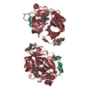
Type: Oligopeptide / Class: Anticoagulant, Antithrombotic / Mass: 1514.605 Da / Num. of mol.: 1 / Source method: obtained synthetically / Details: deduced from C-terminus of hirudin
Source: (natural) Hirudo medicinalis (medicinal leech), (synth.) Synthetic (others)
References: HIRUDIN ANALOGUE
#4: Chemical ChemComp-34P / N-(CARBOXYMETHYL)-3-CYCLOHEXYL-D-ALANYL-N-({4-[(E)-AMINO(IMINO)METHYL]-1,3-THIAZOL-2-YL}METHYL)-L-PROLINAMIDE


Type: peptide-like, Peptide-like / Class: Thrombin inhibitor / Mass: 464.582 Da / Num. of mol.: 1 / Source method: obtained synthetically / Formula: C21H32N6O4S
References: N-(carboxymethyl)-3-cyclohexyl-D-alanyl-N-({4-[(E)-amino(imino)methyl]-1,3-thiazol-2-yl}methyl)-L-prolinamide
#5: Water ChemComp-HOH / water / Water


Mass: 18.015 Da / Num. of mol.: 100 / Source method: isolated from a natural source / Formula: H2O

-
Experimental details

-
Experiment

ExperimentMethod: X-RAY DIFFRACTION / Number of used crystals: 1

-
Sample preparation

CrystalDensity Matthews: 2.61 Å3/Da / Density % sol: 52.9 %
Crystal growTemperature: 298 K / Method: evaporation / pH: 8.5
Details: PEG 8000, Na-acetate, Tris, pH 8.5, EVAPORATION, temperature 298.0K

-
Data collection

DiffractionMean temperature: 100 K
Diffraction sourceSource: ROTATING ANODE / Type: RIGAKU RU200 / Wavelength: 1.5418 Å
DetectorType: SIEMENS HI-STAR / Detector: AREA DETECTOR / Date: Sep 12, 1997 / Details: goebel mirror
RadiationMonochromator: goebel mirror / Protocol: SINGLE WAVELENGTH / Monochromatic (M) / Laue (L): M / Scattering type: x-ray
Radiation wavelengthWavelength: 1.5418 Å / Relative weight: 1
ReflectionResolution: 2.44→50.3 Å / Num. all: 13551 / Num. obs: 12441 / % possible obs: 91.8 % / Observed criterion σ(F): 0 / Observed criterion σ(I): 0 / Redundancy: 3.56 % / Biso Wilson estimate: 37.3 Å2 / Rmerge(I) obs: 0.09 / Rsym value: 0.077 / Net I/σ(I): 10.3
Reflection shellResolution: 2.44→2.59 Å / Redundancy: 1.8 % / Rmerge(I) obs: 0.334 / Mean I/σ(I) obs: 2.3 / Num. unique all: 1208 / Rsym value: 0.249 / % possible all: 54.3

-
Processing

Software
NameClassification
SAINTdata scaling
X-GENdata reduction
CNXrefinement
SAINTdata reduction
X-GENdata scaling
CNXphasing
RefinementMethod to determine structure: MOLECULAR REPLACEMENT / Resolution: 2.44→50.3 Å / Isotropic thermal model: isotropic / σ(F): 0 / Stereochemistry target values: Engh & Huber
RfactorNum. reflection% reflectionSelection details
Rfree0.242 613 -random
Rwork0.185 ---
all0.185 11967 --
obs0.185 11967 5.1 %-
Refine analyze
FreeObs
Luzzati coordinate error0.38 Å0.27 Å
Luzzati d res low-5 Å
Luzzati sigma a0.4 Å0.35 Å
Refinement stepCycle: LAST / Resolution: 2.44→50.3 Å
ProteinNucleic acidLigandSolventTotal
Num. atoms2361 0 32 100 2493
Refine LS restraints
Refine-IDTypeDev ideal
X-RAY DIFFRACTIONc_bond_d0.01
X-RAY DIFFRACTIONc_angle_d1.4
X-RAY DIFFRACTIONc_dihedral_angle_d25.5
X-RAY DIFFRACTIONc_improper_angle_d0.86
LS refinement shellResolution: 2.44→2.59 Å / Rfactor Rfree error: 0.043
RfactorNum. reflection% reflection
Rfree0.317 55 -
Rwork0.28 --
obs-981 0.46 %

+
About Yorodumi

-
News

-
Feb 9, 2022. New format data for meta-information of EMDB entries

New format data for meta-information of EMDB entries

  • Version 3 of the EMDB header file is now the official format.
  • The previous official version 1.9 will be removed from the archive.

Related info.:EMDB header

External links:wwPDB to switch to version 3 of the EMDB data model

-
Aug 12, 2020. Covid-19 info

Covid-19 info

URL: https://pdbj.org/emnavi/covid19.php

New page: Covid-19 featured information page in EM Navigator.

Related info.:Covid-19 info / Mar 5, 2020. Novel coronavirus structure data

+
Mar 5, 2020. Novel coronavirus structure data

Novel coronavirus structure data

Related info.:Yorodumi Speices / Aug 12, 2020. Covid-19 info

External links:COVID-19 featured content - PDBj / Molecule of the Month (242):Coronavirus Proteases

+
Jan 31, 2019. EMDB accession codes are about to change! (news from PDBe EMDB page)

EMDB accession codes are about to change! (news from PDBe EMDB page)

  • The allocation of 4 digits for EMDB accession codes will soon come to an end. Whilst these codes will remain in use, new EMDB accession codes will include an additional digit and will expand incrementally as the available range of codes is exhausted. The current 4-digit format prefixed with “EMD-” (i.e. EMD-XXXX) will advance to a 5-digit format (i.e. EMD-XXXXX), and so on. It is currently estimated that the 4-digit codes will be depleted around Spring 2019, at which point the 5-digit format will come into force.
  • The EM Navigator/Yorodumi systems omit the EMD- prefix.

Related info.:Q: What is EMD? / ID/Accession-code notation in Yorodumi/EM Navigator

External links:EMDB Accession Codes are Changing Soon! / Contact to PDBj

+
Jul 12, 2017. Major update of PDB

Major update of PDB

  • wwPDB released updated PDB data conforming to the new PDBx/mmCIF dictionary.
  • This is a major update changing the version number from 4 to 5, and with Remediation, in which all the entries are updated.
  • In this update, many items about electron microscopy experimental information are reorganized (e.g. em_software).
  • Now, EM Navigator and Yorodumi are based on the updated data.

External links:wwPDB Remediation / Enriched Model Files Conforming to OneDep Data Standards Now Available in the PDB FTP Archive

-
Yorodumi

Thousand views of thousand structures

  • Yorodumi is a browser for structure data from EMDB, PDB, SASBDB, etc.
  • This page is also the successor to EM Navigator detail page, and also detail information page/front-end page for Omokage search.
  • The word "yorodu" (or yorozu) is an old Japanese word meaning "ten thousand". "mi" (miru) is to see.

Related info.:EMDB / PDB / SASBDB / Comparison of 3 databanks / Yorodumi Search / Aug 31, 2016. New EM Navigator & Yorodumi / Yorodumi Papers / Jmol/JSmol / Function and homology information / Changes in new EM Navigator and Yorodumi

Read more