 Movie
Movie Controller

[English] 日本語
 Yorodumi
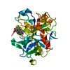
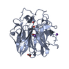
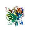
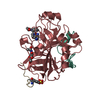
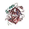
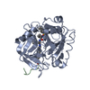
- PDB-5gds: HIRUNORMS ARE TRUE HIRUDIN MIMETICS. THE CRYSTAL STRUCTURE OF HUM... -
+ Open data

- Basic information
| Entry | Database: PDB / ID: 5gds | ||||||
|---|---|---|---|---|---|---|---|
| Title | HIRUNORMS ARE TRUE HIRUDIN MIMETICS. THE CRYSTAL STRUCTURE OF HUMAN ALPHA-THROMBIN:HIRUNORM V COMPLEX | ||||||
 Components Components |
| ||||||
 Keywords Keywords | HYDROLASE/HYDROLASE INHIBITOR / SERINE PROTEASE-INHIBITOR complex / THROMBIN / THROMBIN SYNTHETIC INHIBITORS / ANTITHROMBOTICS / HIRUNORMS / HIRUDIN-LIKE BINDING MODE / BLOOD COAGULATION / HYDROLASE-HYDROLASE INHIBITOR COMPLEX | ||||||
| Function / homology |  Function and homology information Function and homology information: / thrombospondin receptor activity / thrombin / thrombin-activated receptor signaling pathway / Defective factor XII causes hereditary angioedema / negative regulation of astrocyte differentiation / regulation of blood coagulation / neutrophil-mediated killing of gram-negative bacterium / positive regulation of phospholipase C-activating G protein-coupled receptor signaling pathway / Defective F8 cleavage by thrombin ...: / thrombospondin receptor activity / thrombin / thrombin-activated receptor signaling pathway / Defective factor XII causes hereditary angioedema / negative regulation of astrocyte differentiation / regulation of blood coagulation / neutrophil-mediated killing of gram-negative bacterium / positive regulation of phospholipase C-activating G protein-coupled receptor signaling pathway / Defective F8 cleavage by thrombin / ligand-gated ion channel signaling pathway / Platelet Aggregation (Plug Formation) / positive regulation of collagen biosynthetic process / negative regulation of platelet activation / negative regulation of blood coagulation / negative regulation of fibrinolysis / blood coagulation, fibrin clot formation / positive regulation of blood coagulation / Transport of gamma-carboxylated protein precursors from the endoplasmic reticulum to the Golgi apparatus / : / Gamma-carboxylation of protein precursors / Removal of aminoterminal propeptides from gamma-carboxylated proteins / regulation of cytosolic calcium ion concentration / fibrinolysis / : / negative regulation of proteolysis / negative regulation of cytokine production involved in inflammatory response / Regulation of Complement cascade / positive regulation of release of sequestered calcium ion into cytosol / acute-phase response / Cell surface interactions at the vascular wall / Peptide ligand-binding receptors / growth factor activity / positive regulation of receptor signaling pathway via JAK-STAT / lipopolysaccharide binding / platelet activation / positive regulation of protein localization to nucleus / response to wounding / Golgi lumen / Regulation of Insulin-like Growth Factor (IGF) transport and uptake by Insulin-like Growth Factor Binding Proteins (IGFBPs) / positive regulation of reactive oxygen species metabolic process / blood coagulation / positive regulation of insulin secretion / regulation of cell shape / antimicrobial humoral immune response mediated by antimicrobial peptide / heparin binding / Thrombin signalling through proteinase activated receptors (PARs) / positive regulation of cell growth / blood microparticle / G alpha (q) signalling events / positive regulation of phosphatidylinositol 3-kinase/protein kinase B signal transduction / cell surface receptor signaling pathway / endoplasmic reticulum lumen / receptor ligand activity / serine-type endopeptidase activity / signaling receptor binding / calcium ion binding / positive regulation of cell population proliferation / proteolysis / : / extracellular exosome / extracellular region / plasma membrane Similarity search - Function | ||||||
| Biological species |  Homo sapiens (human) Homo sapiens (human) | ||||||
| Method |  X-RAY DIFFRACTION / THE STRUCTURE WAS ANALYZED BY DIFFERENCE FOURIER TECHNIQUES / Resolution: 2.1 Å X-RAY DIFFRACTION / THE STRUCTURE WAS ANALYZED BY DIFFERENCE FOURIER TECHNIQUES / Resolution: 2.1 Å | ||||||
 Authors Authors | De Simone, G. / Lombardi, A. / Galdiero, S. / Nastri, F. / Della Morte, R. / Staiano, N. / Pedone, C. / Bolognesi, M. / Pavone, V. | ||||||
 Citation Citation | Journal: Protein Sci. / Year: 1998 Title: Hirunorms are true hirudin mimetics. The crystal structure of human alpha-thrombin-hirunorm V complex. Authors: De Simone, G. / Lombardi, A. / Galdiero, S. / Nastri, F. / Della Morte, R. / Staiano, N. / Pedone, C. / Bolognesi, M. / Pavone, V. #1: Journal: J.Med.Chem. / Year: 1996Title: Rational Design of True Hirudin Mimetics: Synthesis and Characterization of Multisite-Directed Alpha-Thrombin Inhibitors Authors: Lombardi, A. / Nastri, F. / Della Morte, R. / Rossi, A. / De Rosa, A. / Staiano, N. / Pedone, C. / Pavone, V. #2: Journal: J.Mol.Biol. / Year: 1991Title: Refined Structure of the Hirudin-Thrombin Complex Authors: Rydel, T.J. / Tulinsky, A. / Bode, W. / Huber, R. | ||||||
| History |
|
- Structure visualization
| Structure viewer | Molecule: MolmilJmol/JSmol |
|---|
- Downloads & links
-Download
| PDBx/mmCIF format | 5gds.cif.gz | 80.3 KB | Display | PDBx/mmCIF format |
|---|---|---|---|---|
| PDB format | pdb5gds.ent.gz | 61.3 KB | Display | PDB format |
| PDBx/mmJSON format | 5gds.json.gz | Tree view | PDBx/mmJSON format | |
| Others |  Other downloads Other downloads |
-Validation report
| Arichive directory | https://data.pdbj.org/pub/pdb/validation_reports/gd/5gdsftp://data.pdbj.org/pub/pdb/validation_reports/gd/5gds | HTTPS FTP |
|---|
-Related structure data
| Related structure data | 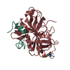 1hahS S: Starting model for refinement |
|---|---|
| Similar structure data |


-Links
 PDBj
PDBj


- Assembly
| Deposited unit | 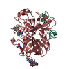
| ||||||||
|---|---|---|---|---|---|---|---|---|---|
| 1 |
| ||||||||
| Unit cell |
|
-Components
| #1: Protein/peptide | Mass: 4096.534 Da / Num. of mol.: 1 / Source method: isolated from a natural source / Source: (natural) Homo sapiens (human) / Organ: BLOOD / Tissue: PLASMA / References: UniProt: P00734, thrombin |
|---|---|
| #2: Protein | Mass: 29780.219 Da / Num. of mol.: 1 / Source method: isolated from a natural source / Source: (natural) Homo sapiens (human) / Organ: BLOOD / Tissue: PLASMA / References: UniProt: P00734, thrombin |
| #3: Protein/peptide | Mass: 2958.211 Da / Num. of mol.: 1 Source method: isolated from a genetically manipulated source |
| #4: Sugar | ChemComp-NAG /   Type: D-saccharide, beta linking / Mass: 221.208 Da / Num. of mol.: 1 Type: D-saccharide, beta linking / Mass: 221.208 Da / Num. of mol.: 1Source method: isolated from a genetically manipulated source Formula: C8H15NO6 |
| #5: Water | ChemComp-HOH /  Mass: 18.015 Da / Num. of mol.: 133 / Source method: isolated from a natural source / Formula: H2O Mass: 18.015 Da / Num. of mol.: 133 / Source method: isolated from a natural source / Formula: H2O |
| Compound details | THROMBIN IS CLEAVED BETWEEN RESIDUES 15 AND 16. CHAIN INDICATOR *L* IS USED FOR RESIDUES 1H - 15 ...THROMBIN IS CLEAVED BETWEEN RESIDUES 15 AND 16. CHAIN INDICATOR *L* IS USED FOR RESIDUES 1H - 15 AND CHAIN INDICATOR *H* IS USED FOR RESIDUES 16 - 247. CHAIN INDICATOR *I* IS USED FOR HIRUNORM V INHIBITOR. |
| Sequence details | CHYMOTRYPSIN NUMBERING (RATHER THAN SEQUENTIAL) SYSTEM IS USED, BASED ON THE TOPOLOGICAL ALIGNMENT ...CHYMOTRYPS |
-Experimental details
-Experiment
| Experiment | Method: X-RAY DIFFRACTION / Number of used crystals: 1 |
|---|
- Sample preparation
| Crystal | Density Matthews: 2.4 Å3/Da / Density % sol: 49 % | ||||||||||||||||||||||||||||||||||||||||||||||||
|---|---|---|---|---|---|---|---|---|---|---|---|---|---|---|---|---|---|---|---|---|---|---|---|---|---|---|---|---|---|---|---|---|---|---|---|---|---|---|---|---|---|---|---|---|---|---|---|---|---|
| Crystal grow | Temperature: 277 K / Method: vapor diffusion / pH: 7 Details: THE CRYSTALS OF THROMBIN:HIRUNORM V COMPLEX WERE GROWN, AS DESCRIBED BY SKRZYPEZAK ET AL. (1991) J. MOL. BIOL.,221,1379-1393 BY VAPOR DIFFUSION METHODS AT 4 C. A 6 MICROLITER DROP, ...Details: THE CRYSTALS OF THROMBIN:HIRUNORM V COMPLEX WERE GROWN, AS DESCRIBED BY SKRZYPEZAK ET AL. (1991) J. MOL. BIOL.,221,1379-1393 BY VAPOR DIFFUSION METHODS AT 4 C. A 6 MICROLITER DROP, CONTAINING 0.05 M SODIUM HEPES (PH 7.0) 10% (W/V) PEG 4000 0.02% NAN3, 20 MG/ML. THROMBIN:HIRUNORM V COMPLEX WAS EQUILIBRATED AGAINST A PRECIPITATING SOLUTION CONTAINING 0.1 M SODIUM HEPES (PH 7.0) 20% (W/V) PEG 4000 0.04% NAN3. CRYSTAL OF THROMBIN:HIRUGEN COMPLEX WERE CRUSHED AND INDIVIDUAL SEEDS WERE USED FOR CROSS-SEEDING EXPERIMENTS., vapor diffusion, temperature 277K | ||||||||||||||||||||||||||||||||||||||||||||||||
| Crystal grow | *PLUS Temperature: 4 ℃ / Method: vapor diffusion | ||||||||||||||||||||||||||||||||||||||||||||||||
| Components of the solutions | *PLUS
|
-Data collection
| Diffraction | Mean temperature: 293 K |
|---|---|
| Diffraction source | Source: ROTATING ANODE / Type: RIGAKU RUH2R / Wavelength: 1.5418 |
| Detector | Type: RIGAKU RAXIS II / Detector: IMAGE PLATE / Date: Jan 10, 1996 / Details: COLLIMATOR |
| Radiation | Monochromatic (M) / Laue (L): M / Scattering type: x-ray |
| Radiation wavelength | Wavelength: 1.5418 Å / Relative weight: 1 |
| Reflection | Resolution: 2.1→20 Å / Num. obs: 21615 / % possible obs: 99.4 % / Redundancy: 2.5 % / Biso Wilson estimate: 28.7 Å2 / Rmerge(I) obs: 0.072 / Net I/σ(I): 5.8 |
| Reflection shell | Resolution: 2.1→2.17 Å / Redundancy: 2.5 % / Rmerge(I) obs: 0.38 / Mean I/σ(I) obs: 2 / % possible all: 98.7 |
| Reflection | *PLUS Num. measured all: 54776 |
| Reflection shell | *PLUS % possible obs: 98.7 % |
- Processing
| Software |
| ||||||||||||||||||||||||||||||||||||||||||||||||||
|---|---|---|---|---|---|---|---|---|---|---|---|---|---|---|---|---|---|---|---|---|---|---|---|---|---|---|---|---|---|---|---|---|---|---|---|---|---|---|---|---|---|---|---|---|---|---|---|---|---|---|---|
| Refinement | Method to determine structure: THE STRUCTURE WAS ANALYZED BY DIFFERENCE FOURIER TECHNIQUES Starting model: PDB ENTRY 1HAH (J. VIJAYALAKSHMI ET AL., 1994, PROTEIN SCI. 3,2254-71) Resolution: 2.1→20 Å / σ(F): 0 Details: THE REGIONS WITH INTERRUPTED ELECTRON DENSITIES ARE FOUND IN THE N-TERMINAL AND C-TERMINAL REGIONS OF CHAIN L, THE C-TERMINAL REGION OF CHAIN H, AND SOME RESIDUES IN THE AUTOLYSIS LOOP. ...Details: THE REGIONS WITH INTERRUPTED ELECTRON DENSITIES ARE FOUND IN THE N-TERMINAL AND C-TERMINAL REGIONS OF CHAIN L, THE C-TERMINAL REGION OF CHAIN H, AND SOME RESIDUES IN THE AUTOLYSIS LOOP. THESE REGIONS INCLUDE: THR L 1H - GLU L 1C; ASP L 14L - ARG L 15; THR H 147 AND TRP H 148; GLY H 246 AND GLU H 247. RESIDUES THR H 149 - LYS H 149E IN THE GAMMA AUTOLYSIS LOOP ARE FOUND WITH NO ELECTRON DENSITIES AND ARE NOT INCLUDED IN THIS ENTRY. THERE IS NO ELECTRON DENSITY FOT HIRUNORM V RESIDUES I 5 - I 15. OTHER ATOMS WITH NO DENSITY ARE GIVEN OCCUPANCY VALUES OF 0.00 IN THIS ENTRY.
| ||||||||||||||||||||||||||||||||||||||||||||||||||
| Solvent computation | Bsol: 158.7 Å2 / ksol: 0.834 e/Å3 | ||||||||||||||||||||||||||||||||||||||||||||||||||
| Refinement step | Cycle: LAST / Resolution: 2.1→20 Å
| ||||||||||||||||||||||||||||||||||||||||||||||||||
| Refine LS restraints |
| ||||||||||||||||||||||||||||||||||||||||||||||||||
| Software | *PLUS Name: TNT / Version: 5E / Classification: refinement | ||||||||||||||||||||||||||||||||||||||||||||||||||
| Refinement | *PLUS Num. reflection obs: 21615 / Rfactor obs: 0.176 | ||||||||||||||||||||||||||||||||||||||||||||||||||
| Solvent computation | *PLUS | ||||||||||||||||||||||||||||||||||||||||||||||||||
| Displacement parameters | *PLUS | ||||||||||||||||||||||||||||||||||||||||||||||||||
| Refine LS restraints | *PLUS
|
