 Movie
Movie Controller

[English] 日本語
 Yorodumi
- PDB-1ook: Crystal Structure of the Complex of Platelet Receptor GPIb-alpha ... -
+ Open data

- Basic information
| Entry | Database: PDB / ID: 1ook | |||||||||
|---|---|---|---|---|---|---|---|---|---|---|
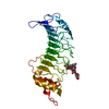
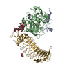
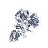
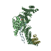
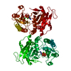
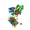
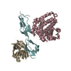
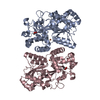
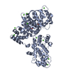
| Title | Crystal Structure of the Complex of Platelet Receptor GPIb-alpha and Human alpha-Thrombin | |||||||||
 Components Components |
| |||||||||
 Keywords Keywords | HYDROLASE / Leucine rich repeats | |||||||||
| Function / homology |  Function and homology information Function and homology informationthrombin-activated receptor activity / glycoprotein Ib-IX-V complex / Enhanced binding of GP1BA variant to VWF multimer:collagen / Defective binding of VWF variant to GPIb:IX:V / blood coagulation, intrinsic pathway / Defective F9 activation / Platelet Adhesion to exposed collagen / positive regulation of platelet activation / megakaryocyte development / : ...thrombin-activated receptor activity / glycoprotein Ib-IX-V complex / Enhanced binding of GP1BA variant to VWF multimer:collagen / Defective binding of VWF variant to GPIb:IX:V / blood coagulation, intrinsic pathway / Defective F9 activation / Platelet Adhesion to exposed collagen / positive regulation of platelet activation / megakaryocyte development / : / GP1b-IX-V activation signalling / thrombospondin receptor activity / thrombin / thrombin-activated receptor signaling pathway / Defective factor XII causes hereditary angioedema / negative regulation of astrocyte differentiation / regulation of blood coagulation / neutrophil-mediated killing of gram-negative bacterium / positive regulation of phospholipase C-activating G protein-coupled receptor signaling pathway / Defective F8 cleavage by thrombin / ligand-gated ion channel signaling pathway / Platelet Aggregation (Plug Formation) / positive regulation of collagen biosynthetic process / negative regulation of platelet activation / negative regulation of blood coagulation / negative regulation of fibrinolysis / blood coagulation, fibrin clot formation / positive regulation of blood coagulation / Transport of gamma-carboxylated protein precursors from the endoplasmic reticulum to the Golgi apparatus / : / Gamma-carboxylation of protein precursors / Removal of aminoterminal propeptides from gamma-carboxylated proteins / regulation of cytosolic calcium ion concentration / fibrinolysis / : / negative regulation of proteolysis / release of sequestered calcium ion into cytosol / negative regulation of cytokine production involved in inflammatory response / Regulation of Complement cascade / acute-phase response / Cell surface interactions at the vascular wall / positive regulation of release of sequestered calcium ion into cytosol / Peptide ligand-binding receptors / growth factor activity / positive regulation of receptor signaling pathway via JAK-STAT / lipopolysaccharide binding / RUNX1 regulates genes involved in megakaryocyte differentiation and platelet function / platelet activation / response to wounding / positive regulation of protein localization to nucleus / Golgi lumen / Regulation of Insulin-like Growth Factor (IGF) transport and uptake by Insulin-like Growth Factor Binding Proteins (IGFBPs) / cell morphogenesis / positive regulation of reactive oxygen species metabolic process / blood coagulation / positive regulation of insulin secretion / regulation of cell shape / antimicrobial humoral immune response mediated by antimicrobial peptide / heparin binding / Thrombin signalling through proteinase activated receptors (PARs) / positive regulation of cell growth / signaling receptor activity / blood microparticle / G alpha (q) signalling events / cell surface receptor signaling pathway / positive regulation of phosphatidylinositol 3-kinase/protein kinase B signal transduction / cell adhesion / endoplasmic reticulum lumen / receptor ligand activity / signaling receptor binding / serine-type endopeptidase activity / external side of plasma membrane / calcium ion binding / positive regulation of cell population proliferation / cell surface / proteolysis / : / extracellular exosome / extracellular region / membrane / plasma membrane Similarity search - Function | |||||||||
| Biological species |  Homo sapiens (human) Homo sapiens (human) | |||||||||
| Method |  X-RAY DIFFRACTION / SYNCHROTRON / MOLECULAR REPLACEMENT / Resolution: 2.3 Å X-RAY DIFFRACTION / SYNCHROTRON / MOLECULAR REPLACEMENT / Resolution: 2.3 Å | |||||||||
 Authors Authors | Varughese, K.I. / Celikel, R. / Ruggeri, Z.M. | |||||||||
 Citation Citation | Journal: Science / Year: 2003 Title: Modulation of alpha-thrombin function by distinct interactions with platelet glycoprotein Ibalpha Authors: Celikel, R. / McClintock, R.A. / Roberts, J.R. / Mendolicchio, G.L. / Ware, J. / Varughese, K.I. / Ruggeri, Z.M. | |||||||||
| History |
|
- Structure visualization
| Structure viewer | Molecule: MolmilJmol/JSmol |
|---|
- Downloads & links
-Download
| PDBx/mmCIF format | 1ook.cif.gz | 144 KB | Display | PDBx/mmCIF format |
|---|---|---|---|---|
| PDB format | pdb1ook.ent.gz | 109.8 KB | Display | PDB format |
| PDBx/mmJSON format | 1ook.json.gz | Tree view | PDBx/mmJSON format | |
| Others |  Other downloads Other downloads |
-Validation report
| Arichive directory | https://data.pdbj.org/pub/pdb/validation_reports/oo/1ookftp://data.pdbj.org/pub/pdb/validation_reports/oo/1ook | HTTPS FTP |
|---|
-Related structure data
-Links
 PDBj
PDBj


- Assembly
| Deposited unit | 
| ||||||||
|---|---|---|---|---|---|---|---|---|---|
| 1 |
| ||||||||
| Unit cell |
|
-Components
-Protein/peptide , 2 types, 2 molecules AP
| #1: Protein/peptide | Mass: 4096.534 Da / Num. of mol.: 1 / Fragment: THROMBIN A CHAIN / Source method: isolated from a natural source / Details: Purified from plasma / Source: (natural) Homo sapiens (human) / References: UniProt: P00734, thrombin |
|---|---|
| #3: Protein/peptide | Mass: 419.498 Da / Num. of mol.: 1 / Source method: obtained synthetically / Details: Chemically synthesized |
-Protein , 2 types, 2 molecules BG
| #2: Protein | Mass: 29780.219 Da / Num. of mol.: 1 / Fragment: THROMBIN B CHAIN / Source method: isolated from a natural source / Details: Purified from plasma / Source: (natural) Homo sapiens (human) / References: UniProt: P00734, thrombin |
|---|---|
| #4: Protein | Mass: 32466.920 Da / Num. of mol.: 1 / Fragment: N-terminal domain / Mutation: C65A Source method: isolated from a genetically manipulated source Source: (gene. exp.) Homo sapiens (human) / Gene: GP1BA / Production host:  |
-Sugars , 2 types, 2 molecules 
| #5: Polysaccharide | alpha-D-mannopyranose-(1-3)-[alpha-D-mannopyranose-(1-6)]alpha-D-mannopyranose-(1-4)-2-acetamido-2- ...alpha-D-mannopyranose-(1-3)-[alpha-D-mannopyranose-(1-6)]alpha-D-mannopyranose-(1-4)-2-acetamido-2-deoxy-beta-D-glucopyranose-(1-4)-2-acetamido-2-deoxy-beta-D-glucopyranose Source method: isolated from a genetically manipulated source |
|---|---|
| #7: Sugar | ChemComp-NAG /  Type: D-saccharide, beta linking / Mass: 221.208 Da / Num. of mol.: 1 Type: D-saccharide, beta linking / Mass: 221.208 Da / Num. of mol.: 1Source method: isolated from a genetically manipulated source Formula: C8H15NO6 |
-Non-polymers , 2 types, 393 molecules 

| #6: Chemical | ChemComp-CL /  Mass: 35.453 Da / Num. of mol.: 1 / Source method: obtained synthetically / Formula: Cl Mass: 35.453 Da / Num. of mol.: 1 / Source method: obtained synthetically / Formula: Cl |
|---|---|
| #8: Water | ChemComp-HOH / Mass: 18.015 Da / Num. of mol.: 392 / Source method: isolated from a natural source / Formula: H2O |
-Details
| Has protein modification | Y |
|---|
-Experimental details
-Experiment
| Experiment | Method: X-RAY DIFFRACTION / Number of used crystals: 1 |
|---|
- Sample preparation
| Crystal | Density Matthews: 2.63 Å3/Da / Density % sol: 52.81 % | ||||||||||||||||||||||||||||||||||||||||||
|---|---|---|---|---|---|---|---|---|---|---|---|---|---|---|---|---|---|---|---|---|---|---|---|---|---|---|---|---|---|---|---|---|---|---|---|---|---|---|---|---|---|---|---|
| Crystal grow | Temperature: 277 K / Method: vapor diffusion, hanging drop / pH: 7 Details: PEG 6000, Ammonium Phosphate, pH 7.0, VAPOR DIFFUSION, HANGING DROP, temperature 277.0K | ||||||||||||||||||||||||||||||||||||||||||
| Crystal grow | *PLUS Temperature: 4 ℃ / pH: 7.4 / Method: vapor diffusion, hanging drop | ||||||||||||||||||||||||||||||||||||||||||
| Components of the solutions | *PLUS
|
-Data collection
| Diffraction | Mean temperature: 100 K |
|---|---|
| Diffraction source | Source: SYNCHROTRON / Site: SSRL  / Beamline: BL9-2 / Wavelength: 1.033167 Å / Beamline: BL9-2 / Wavelength: 1.033167 Å |
| Detector | Type: ADSC QUANTUM 4 / Detector: CCD / Date: Dec 2, 2002 |
| Radiation | Monochromator: Double Crystal / Protocol: SINGLE WAVELENGTH / Monochromatic (M) / Laue (L): M / Scattering type: x-ray |
| Radiation wavelength | Wavelength: 1.033167 Å / Relative weight: 1 |
| Reflection | Resolution: 2.3→50 Å / Num. all: 35360 / Num. obs: 35360 / % possible obs: 99.7 % / Observed criterion σ(F): 0 / Observed criterion σ(I): 0 / Redundancy: 17.2 % / Biso Wilson estimate: 31.9 Å2 / Rsym value: 0.066 / Net I/σ(I): 38.6 |
| Reflection shell | Resolution: 2.3→2.38 Å / Mean I/σ(I) obs: 5.2 / Rsym value: 0.292 / % possible all: 97.5 |
| Reflection | *PLUS Num. measured all: 608108 / Rmerge(I) obs: 0.066 |
| Reflection shell | *PLUS % possible obs: 97.5 % / Rmerge(I) obs: 0.292 |
- Processing
| Software |
| |||||||||||||||||||||||||
|---|---|---|---|---|---|---|---|---|---|---|---|---|---|---|---|---|---|---|---|---|---|---|---|---|---|---|
| Refinement | Method to determine structure: MOLECULAR REPLACEMENT / Resolution: 2.3→15 Å / σ(F): 0 / σ(I): 0 / Stereochemistry target values: Engh & Huber
| |||||||||||||||||||||||||
| Refine analyze | Luzzati coordinate error obs: 0.28 Å / Luzzati d res low obs: 5 Å / Luzzati sigma a obs: 0.24 Å | |||||||||||||||||||||||||
| Refinement step | Cycle: LAST / Resolution: 2.3→15 Å
| |||||||||||||||||||||||||
| Refine LS restraints |
| |||||||||||||||||||||||||
| Refinement | *PLUS Highest resolution: 2.3 Å / Lowest resolution: 15 Å / Num. reflection obs: 32958 / Num. reflection Rfree: 1722 / Rfactor Rfree: 0.26 | |||||||||||||||||||||||||
| Solvent computation | *PLUS | |||||||||||||||||||||||||
| Displacement parameters | *PLUS | |||||||||||||||||||||||||
| Refine LS restraints | *PLUS
| |||||||||||||||||||||||||
| LS refinement shell | *PLUS Rfactor Rfree: 0.345 / Rfactor Rwork: 0.2831 |
