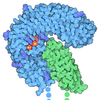
Entry Database : PDB / ID : 3pmhTitle Mechanism of Sulfotyrosine-Mediated Glycoprotein Ib Interaction with Two Distinct alpha-Thrombin Sites Platelet glycoprotein Ib alpha chain THROMBIN ALPHA-CHAIN THROMBIN BETA-CHAIN Keywords / / / / / / / Function / homology Function Domain/homology Component
/ / / / / / / / / / / / / / / / / / / / / / / / / / / / / / / / / / / / / / / / / / / / / / / / / / / / / / / / / / / / / / / / / / / / / / / / / / / / / / / / / / / / / / / / / / / / / / / / / / / / / / / / / / / / / / / / / / / / / / / / / / / / / / / / / / / / / / / / / / / / / / / / / / / Biological species Homo sapiens (human)Method / / / Resolution : 3.2 Å Authors Varughese, K.I. / Celikel, R. Journal : Proc.Natl.Acad.Sci.USA / Year : 2011Title : Binding of alpha-thrombin to surface-anchored platelet glycoprotein Ib(alpha) sulfotyrosines through a two-site mechanism involving exosite I.Authors : Zarpellon, A. / Celikel, R. / Roberts, J.R. / McClintock, R.A. / Mendolicchio, G.L. / Moore, K.L. / Jing, H. / Varughese, K.I. / Ruggeri, Z.M. History Deposition Nov 16, 2010 Deposition site / Processing site Revision 1.0 Jun 1, 2011 Provider / Type Revision 1.1 Jul 13, 2011 Group Revision 1.2 Aug 10, 2011 Group Revision 1.3 Dec 12, 2012 Group Revision 2.0 Jul 29, 2020 Group Atomic model / Data collection ... Atomic model / Data collection / Database references / Derived calculations / Structure summary Category atom_site / chem_comp ... atom_site / chem_comp / entity / entity_name_com / pdbx_branch_scheme / pdbx_chem_comp_identifier / pdbx_entity_branch / pdbx_entity_branch_descriptor / pdbx_entity_branch_link / pdbx_entity_branch_list / pdbx_entity_nonpoly / pdbx_molecule / pdbx_nonpoly_scheme / pdbx_struct_assembly_gen / struct_asym / struct_conn / struct_ref_seq_dif / struct_site / struct_site_gen Item _atom_site.B_iso_or_equiv / _atom_site.Cartn_x ... _atom_site.B_iso_or_equiv / _atom_site.Cartn_x / _atom_site.Cartn_y / _atom_site.Cartn_z / _atom_site.auth_asym_id / _atom_site.auth_atom_id / _atom_site.auth_comp_id / _atom_site.auth_seq_id / _atom_site.label_asym_id / _atom_site.label_atom_id / _atom_site.label_comp_id / _atom_site.label_entity_id / _atom_site.type_symbol / _chem_comp.name / _chem_comp.type / _entity.formula_weight / _entity.pdbx_description / _entity.pdbx_number_of_molecules / _entity.src_method / _entity.type / _entity_name_com.entity_id / _pdbx_molecule.asym_id / _pdbx_struct_assembly_gen.asym_id_list / _struct_conn.pdbx_dist_value / _struct_conn.pdbx_leaving_atom_flag / _struct_conn.pdbx_ptnr1_PDB_ins_code / _struct_conn.pdbx_role / _struct_conn.ptnr1_auth_asym_id / _struct_conn.ptnr1_auth_comp_id / _struct_conn.ptnr1_auth_seq_id / _struct_conn.ptnr1_label_asym_id / _struct_conn.ptnr1_label_atom_id / _struct_conn.ptnr1_label_comp_id / _struct_conn.ptnr1_label_seq_id / _struct_conn.ptnr2_auth_asym_id / _struct_conn.ptnr2_auth_comp_id / _struct_conn.ptnr2_auth_seq_id / _struct_conn.ptnr2_label_asym_id / _struct_conn.ptnr2_label_atom_id / _struct_conn.ptnr2_label_comp_id / _struct_conn.ptnr2_label_seq_id / _struct_ref_seq_dif.details Description / Provider / Type Revision 2.1 Sep 6, 2023 Group Data collection / Database references ... Data collection / Database references / Refinement description / Structure summary Category chem_comp / chem_comp_atom ... chem_comp / chem_comp_atom / chem_comp_bond / database_2 / pdbx_initial_refinement_model Item / _database_2.pdbx_DOI / _database_2.pdbx_database_accessionRevision 2.2 Dec 6, 2023 Group / Category / chem_comp_bond / Item / _chem_comp_bond.atom_id_2Revision 2.3 Oct 30, 2024 Group / Category / pdbx_modification_feature
Show all Show less
 Yorodumi
 Open data
 Basic information
 Components
 Keywords
 Function and homology information
 Homo sapiens (human)
 X-RAY DIFFRACTION /
  SYNCHROTRON /
  MOLECULAR REPLACEMENT / Resolution: 3.2 Å
  Authors
 Citation
 Journal: Proc.Natl.Acad.Sci.USA / Year: 2011
 Structure visualization
 Molmil
 Jmol/JSmol
 Downloads & links
 Download
 3pmh.cif.gz
 PDBx/mmCIF format
 pdb3pmh.ent.gz
 PDB format
 3pmh.json.gz
 PDBx/mmJSON format
 Other downloads
 https://data.pdbj.org/pub/pdb/validation_reports/pm/3pmh
 ftp://data.pdbj.org/pub/pdb/validation_reports/pm/3pmh
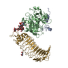
 Links
Links Assembly
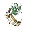
 Components
 Homo sapiens (human) / References: UniProt: P00734, thrombin
 Homo sapiens (human) / Gene: GP1BA, platelet glycoprotein Ib alpha / Production host:
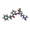

 Homo sapiens (human) / References: UniProt: P00734, thrombin
 X-RAY DIFFRACTION / Number of used crystals: 1
  Sample preparation
 SYNCHROTRON / Site:
  SSRL
SSRL  / Beamline: BL9-2 / Wavelength: 1.0332
  Processing
 MOLECULAR REPLACEMENT
 Movie
Movie Controller


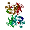
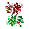
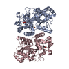
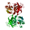
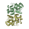
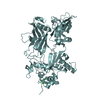
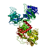

 PDBj
PDBj


