 Movie
Movie Controller


+ Open data

- Basic information
| Entry | Database: PDB / ID: 1cb6 | ||||||
|---|---|---|---|---|---|---|---|
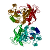
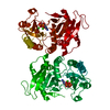
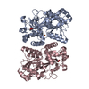
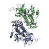
| Title | STRUCTURE OF HUMAN APOLACTOFERRIN AT 2.0 A RESOLUTION. | ||||||
 Components Components | Lactotransferrin | ||||||
 Keywords Keywords | IRON TRANSPORT / APOLACTOFERRIN / CONFORMATIONAL CHANGE | ||||||
| Function / homology |  Function and homology information Function and homology informationmembrane destabilizing activity / host-mediated suppression of viral proces / Mtb iron assimilation by chelation / phagocytic vesicle lumen / Metal sequestration by antimicrobial proteins / negative regulation of viral process / negative regulation of tumor necrosis factor (ligand) superfamily member 11 production / positive regulation of toll-like receptor 4 signaling pathway / negative regulation of single-species biofilm formation in or on host organism / positive regulation of bone mineralization involved in bone maturation ...membrane destabilizing activity / host-mediated suppression of viral proces / Mtb iron assimilation by chelation / phagocytic vesicle lumen / Metal sequestration by antimicrobial proteins / negative regulation of viral process / negative regulation of tumor necrosis factor (ligand) superfamily member 11 production / positive regulation of toll-like receptor 4 signaling pathway / negative regulation of single-species biofilm formation in or on host organism / positive regulation of bone mineralization involved in bone maturation / negative regulation of ATP-dependent activity / negative regulation of osteoclast development / specific granule / negative regulation of lipopolysaccharide-mediated signaling pathway / positive regulation of chondrocyte proliferation / antifungal humoral response / regulation of tumor necrosis factor production / bone morphogenesis / Antimicrobial peptides / positive regulation of osteoblast proliferation / negative regulation of viral genome replication / positive regulation of protein serine/threonine kinase activity / humoral immune response / Hydrolases; Acting on peptide bonds (peptidases); Serine endopeptidases / positive regulation of osteoblast differentiation / cysteine-type endopeptidase inhibitor activity / : / regulation of cytokine production / secretory granule / ossification / protein serine/threonine kinase activator activity / innate immune response in mucosa / iron ion transport / lipopolysaccharide binding / recycling endosome / specific granule lumen / tertiary granule lumen / antimicrobial humoral immune response mediated by antimicrobial peptide / heparin binding / antibacterial humoral response / killing of cells of another organism / defense response to Gram-negative bacterium / early endosome / positive regulation of canonical NF-kappaB signal transduction / iron ion binding / Amyloid fiber formation / serine-type endopeptidase activity / Neutrophil degranulation / negative regulation of apoptotic process / cell surface / protein-containing complex / proteolysis / : / DNA binding / extracellular exosome / extracellular region / nucleus / plasma membrane / cytoplasm Similarity search - Function | ||||||
| Biological species |  Homo sapiens (human) Homo sapiens (human) | ||||||
| Method |  X-RAY DIFFRACTION / SYNCHROTRON / MOLECULAR REPLACEMENT / Resolution: 2 Å X-RAY DIFFRACTION / SYNCHROTRON / MOLECULAR REPLACEMENT / Resolution: 2 Å | ||||||
 Authors Authors | Jameson, G.B. / Anderson, B.F. / Norris, G.E. / Thomas, D.H. / Baker, E.N. | ||||||
 Citation Citation | Journal: Acta Crystallogr.,Sect.D / Year: 1998 Title: Structure of human apolactoferrin at 2.0 A resolution. Refinement and analysis of ligand-induced conformational change. Authors: Jameson, G.B. / Anderson, B.F. / Norris, G.E. / Thomas, D.H. / Baker, E.N. #1: Journal: Nature / Year: 1990 Title: Apolactoferrin structure demonstrates ligand-induced conformational change in transferrins. Authors: Anderson, B.F. / Baker, H.M. / Norris, G.E. / Rumball, S.V. / Baker, E.N. #2: Journal: J.Mol.Biol. / Year: 1989 Title: Structure of human lactoferrin: crystallographic structure analysis and refinement at 2.8 A resolution. Authors: Anderson, B.F. / Baker, H.M. / Norris, G.E. / Rice, D.W. / Baker, E.N. #3: Journal: Trends Biochem.Sci. / Year: 1987Title: Transferrins: Insights Into Structure and Function from Studies on Lactoferrin Authors: Baker, E.N. / Rumball, S.V. / Anderson, B.F. #4: Journal: Proc.Natl.Acad.Sci.USA / Year: 1987 Title: Structure of human lactoferrin at 3.2-A resolution. Authors: Anderson, B.F. / Baker, H.M. / Dodson, E.J. / Norris, G.E. / Rumball, S.V. / Waters, J.M. / Baker, E.N. | ||||||
| History |
| ||||||
| Remark 650 | HELIX DETERMINATION METHOD: AUTHOR-MODIFIED KABSCH & SANDER | ||||||
| Remark 700 | SHEET THERE ARE SEVERAL BIFURCATED SHEETS IN THIS STRUCTURE. THESE ARE REPRESENTED BY TWO SHEETS ...SHEET THERE ARE SEVERAL BIFURCATED SHEETS IN THIS STRUCTURE. THESE ARE REPRESENTED BY TWO SHEETS WHICH HAVE ONE OR MORE IDENTICAL STRANDS. SHEETS *N2A* AND *N2B* REPRESENT ONE BIFURCATED SHEET AND STRAND FOUR OF SHEET BN1 IS ALSO PART OF THIS SHEET. SHEETS *C2A* AND *C2B* REPRESENT ONE BIFURCATED SHEET AND STRAND FOUR OF SHEET BC1 IS ALSO PART OF THIS SHEET. THE RESIDUES LISTED IN REMARK 500, TORSION ANGLES OUTSIDE THE EXPECTED RAMACHANDRAN REGIONS, ARE IN WELL DEFINED GAMMA-TURNS AT POSITIONALLY HOMOLOGOUS SITES ON BOTH LOBES. THE PEPTIDE BOND IN REMARK 500 AND ITS POSITIONAL HOMOLOG ARE ALSO SUBSTANTIALLY NON-PLANAR IN LACTOFERRIN |
- Structure visualization
| Structure viewer | Molecule: MolmilJmol/JSmol |
|---|
- Downloads & links
-Download
| PDBx/mmCIF format | 1cb6.cif.gz | 156.3 KB | Display | PDBx/mmCIF format |
|---|---|---|---|---|
| PDB format | pdb1cb6.ent.gz | 121.2 KB | Display | PDB format |
| PDBx/mmJSON format | 1cb6.json.gz | Tree view | PDBx/mmJSON format | |
| Others |  Other downloads Other downloads |
-Validation report
| Arichive directory | https://data.pdbj.org/pub/pdb/validation_reports/cb/1cb6ftp://data.pdbj.org/pub/pdb/validation_reports/cb/1cb6 | HTTPS FTP |
|---|
-Related structure data
| Related structure data |  1lgfS S: Starting model for refinement |
|---|---|
| Similar structure data |
-Links
 PDBj
PDBj


- Assembly
| Deposited unit | 
| ||||||||
|---|---|---|---|---|---|---|---|---|---|
| 1 |
| ||||||||
| Unit cell |
|
-Components
| #1: Protein | Mass: 76263.266 Da / Num. of mol.: 1 / Fragment: UNP residues 20-710 / Source method: isolated from a natural source / Source: (natural) Homo sapiens (human) / Organ: BREAST / Secretion: MILKReferences: UniProt: P02788, Hydrolases; Acting on peptide bonds (peptidases); Serine endopeptidases | ||||||||
|---|---|---|---|---|---|---|---|---|---|
| #2: Chemical |   Mass: 35.453 Da / Num. of mol.: 2 / Source method: obtained synthetically / Formula: Cl Mass: 35.453 Da / Num. of mol.: 2 / Source method: obtained synthetically / Formula: Cl#3: Water | ChemComp-HOH / |  Mass: 18.015 Da / Num. of mol.: 357 / Source method: isolated from a natural source / Formula: H2O Mass: 18.015 Da / Num. of mol.: 357 / Source method: isolated from a natural source / Formula: H2OCompound details | THERE IS A TWO FOLD INTERNAL SEQUENCE HOMOLOGY (~40% IDENTITY). | Has protein modification | Y | Sequence details | THE SEQUENCE USED IN THE X-RAY STRUCTURE FOR RESIDUES 1001-1005 (GRRRS) IS AT VARIANCE WITH THE ...THE SEQUENCE USED IN THE X-RAY STRUCTURE FOR RESIDUES 1001-1005 (GRRRS) IS AT VARIANCE WITH THE MOST RECENT SEQUENCE (SWS P02788, TRFL_H: GRRRRS). IN THIS REGION ELECTRON DENSITY IS POORLY DEFINED AND THE DIFFERENCE | |
-Experimental details
-Experiment
| Experiment | Method: X-RAY DIFFRACTION / Number of used crystals: 3 |
|---|
- Sample preparation
| Crystal | Density Matthews: 2.61 Å3/Da / Density % sol: 53 % Description: SYNCHROTRON OSCILLATION DATA TO 1.9 A MERGED WITH ENRAF-NONIUS CAD4 DATA TO 2.8 A; DATA TO 2.0 A RETAINED | |||||||||||||||||||||||||
|---|---|---|---|---|---|---|---|---|---|---|---|---|---|---|---|---|---|---|---|---|---|---|---|---|---|---|
| Crystal grow | pH: 7.8 / Details: pH 7.8 | |||||||||||||||||||||||||
| Crystal grow | *PLUS Method: microdialysis | |||||||||||||||||||||||||
| Components of the solutions | *PLUS
|
-Data collection
| Diffraction | Mean temperature: 295 K |
|---|---|
| Diffraction source | Source: SYNCHROTRON / Site: SRS  / Type: SRS / Wavelength: 0.87 / Type: SRS / Wavelength: 0.87 |
| Detector | Detector: FILM / Details: MIRRORS |
| Radiation | Monochromator: NI MIRRORS / Protocol: SINGLE WAVELENGTH / Monochromatic (M) / Laue (L): M / Scattering type: x-ray |
| Radiation wavelength | Wavelength: 0.87 Å / Relative weight: 1 |
| Reflection | Resolution: 2→17 Å / Num. obs: 53450 / % possible obs: 92 % / Redundancy: 3.3 % / Biso Wilson estimate: 42.3 Å2 / Rmerge(I) obs: 0.083 / Net I/σ(I): 30.3 |
| Reflection shell | Resolution: 2→2.12 Å / Redundancy: 3 % / Rmerge(I) obs: 0.61 / Mean I/σ(I) obs: 2.7 / % possible all: 91 |
| Reflection shell | *PLUS % possible obs: 91 % |
- Processing
| Software |
| |||||||||||||||||||||||||||||||||
|---|---|---|---|---|---|---|---|---|---|---|---|---|---|---|---|---|---|---|---|---|---|---|---|---|---|---|---|---|---|---|---|---|---|---|
| Refinement | Method to determine structure: MOLECULAR REPLACEMENT Starting model: 1LGF (LACTOFERRIN) Resolution: 2→10 Å / Num. parameters: 22947 / Num. restraintsaints: 22209 / Cross valid method: THROUGHOUT / σ(F): 0 Details: ANISOTROPIC SCALING APPLIED BY THE METHOD OF PARKIN, MOEZZI & HOPE, J.APPL.CRYST.28(1995)53-56. PROLSQ WAS USED FOR INITIAL REFINEMENTS. SHELXL97 WAS USED FOR FINAL REFINEMENTS. THERE IS ...Details: ANISOTROPIC SCALING APPLIED BY THE METHOD OF PARKIN, MOEZZI & HOPE, J.APPL.CRYST.28(1995)53-56. PROLSQ WAS USED FOR INITIAL REFINEMENTS. SHELXL97 WAS USED FOR FINAL REFINEMENTS. THERE IS POOR DENSITY FOR RESIDUES 1-3. DENSITY FOR RESIDUES 418 - 424 IS POORLY DEFINED.
| |||||||||||||||||||||||||||||||||
| Solvent computation | Solvent model: MOEWS & KRETSINGER, J.MOL.BIOL.91(1973)201-228 | |||||||||||||||||||||||||||||||||
| Displacement parameters | Biso mean: 53.5 Å2 | |||||||||||||||||||||||||||||||||
| Refinement step | Cycle: LAST / Resolution: 2→10 Å
| |||||||||||||||||||||||||||||||||
| Refine LS restraints |
| |||||||||||||||||||||||||||||||||
| Software | *PLUS Name: SHELXL-97 / Classification: refinement | |||||||||||||||||||||||||||||||||
| Refine LS restraints | *PLUS
|
