 Movie
Movie Controller

[English] 日本語
 Yorodumi
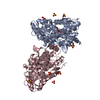
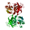
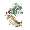
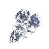
- PDB-3osq: Maltose-bound maltose sensor engineered by insertion of circularl... -
+ Open data

- Basic information
| Entry | Database: PDB / ID: 3osq | |||||||||||||||
|---|---|---|---|---|---|---|---|---|---|---|---|---|---|---|---|---|
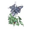
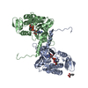
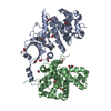
| Title | Maltose-bound maltose sensor engineered by insertion of circularly permuted green fluorescent protein into E. coli maltose binding protein at position 175 | |||||||||||||||
 Components Components | Maltose-binding periplasmic protein,Green fluorescent protein | |||||||||||||||
 Keywords Keywords | FLUORESCENT PROTEIN / Transport Protein / Engineered Protein / Sensor Protein / MBP / GFP / Maltose Sensor | |||||||||||||||
| Function / homology |  Function and homology information Function and homology informationcarbohydrate transmembrane transporter activity / maltose binding / maltose transport / maltodextrin transmembrane transport / ATP-binding cassette (ABC) transporter complex, substrate-binding subunit-containing / bioluminescence / generation of precursor metabolites and energy / outer membrane-bounded periplasmic space Similarity search - Function | |||||||||||||||
| Biological species |    Aequorea victoria (jellyfish) Aequorea victoria (jellyfish) | |||||||||||||||
| Method |  X-RAY DIFFRACTION / SYNCHROTRON / MOLECULAR REPLACEMENT / Resolution: 1.9 Å X-RAY DIFFRACTION / SYNCHROTRON / MOLECULAR REPLACEMENT / Resolution: 1.9 Å | |||||||||||||||
 Authors Authors | Echevarria, I.M. / Marvin, J.S. / Looger, L.L. / Schreiter, E.R. | |||||||||||||||
 Citation Citation | Journal: Proteins / Year: 2011 Title: A genetically encoded, high-signal-to-noise maltose sensor. Authors: Marvin, J.S. / Schreiter, E.R. / Echevarria, I.M. / Looger, L.L. | |||||||||||||||
| History |
|
- Structure visualization
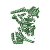
| Structure viewer | Molecule: MolmilJmol/JSmol |
|---|
- Downloads & links
-Download
| PDBx/mmCIF format | 3osq.cif.gz | 258 KB | Display | PDBx/mmCIF format |
|---|---|---|---|---|
| PDB format | pdb3osq.ent.gz | 204.5 KB | Display | PDB format |
| PDBx/mmJSON format | 3osq.json.gz | Tree view | PDBx/mmJSON format | |
| Others |  Other downloads Other downloads |
-Validation report
| Arichive directory | https://data.pdbj.org/pub/pdb/validation_reports/os/3osqftp://data.pdbj.org/pub/pdb/validation_reports/os/3osq | HTTPS FTP |
|---|
-Related structure data
| Related structure data | 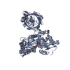 3osrC  1anfS 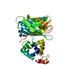 3ek4S C: citing same article ( S: Starting model for refinement |
|---|---|
| Similar structure data |


-Links
 PDBj
PDBj


- Assembly
| Deposited unit | 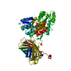
| ||||||||
|---|---|---|---|---|---|---|---|---|---|
| 1 |
| ||||||||
| Unit cell |
|
-Components
| #1: Protein | Mass: 73705.000 Da / Num. of mol.: 1 Fragment: GFP P42212 residues 2-146, 147-238, MBP P0AEX9 residues 27-199, 201-396 Source method: isolated from a genetically manipulated source Source: (gene. exp.) Aequorea victoria (jellyfish)Gene: malE, Z5632, ECs5017, GFP / Plasmid: pRSET / Production host: | ||||
|---|---|---|---|---|---|
| #2: Polysaccharide | alpha-D-glucopyranose-(1-4)-alpha-D-glucopyranose / alpha-maltose  Source method: isolated from a genetically manipulated source Details: oligosaccharide / References: alpha-maltose | ||||
| #3: Chemical |   Mass: 96.063 Da / Num. of mol.: 2 / Source method: obtained synthetically / Formula: SO4 Mass: 96.063 Da / Num. of mol.: 2 / Source method: obtained synthetically / Formula: SO4#4: Water | ChemComp-HOH / |  Mass: 18.015 Da / Num. of mol.: 294 / Source method: isolated from a natural source / Formula: H2O Mass: 18.015 Da / Num. of mol.: 294 / Source method: isolated from a natural source / Formula: H2OHas protein modification | Y | |
-Experimental details
-Experiment
| Experiment | Method: X-RAY DIFFRACTION / Number of used crystals: 1 |
|---|
- Sample preparation
| Crystal | Density Matthews: 2.92 Å3/Da / Density % sol: 57.84 % |
|---|---|
| Crystal grow | Temperature: 296 K / Method: vapor diffusion, hanging drop / pH: 5.6 Details: 0.5 M Ammonium sulfate, 0.1M Sodium citrate tribasic dihydrate pH 5.6, 1.0 M Lithium sulfate monohydrate, VAPOR DIFFUSION, HANGING DROP, temperature 296K |
-Data collection
| Diffraction | Mean temperature: 100 K |
|---|---|
| Diffraction source | Source: SYNCHROTRON / Site: APS  / Beamline: 31-ID / Wavelength: 0.9793 Å / Beamline: 31-ID / Wavelength: 0.9793 Å |
| Detector | Type: RAYONIX MX225HE / Detector: CCD / Date: Jul 21, 2010 |
| Radiation | Monochromator: Diamond (111) double crystal / Protocol: SINGLE WAVELENGTH / Monochromatic (M) / Laue (L): M / Scattering type: x-ray |
| Radiation wavelength | Wavelength: 0.9793 Å / Relative weight: 1 |
| Reflection | Resolution: 1.9→31.41 Å / Num. obs: 68612 / % possible obs: 99.9 % / Observed criterion σ(F): 0 / Observed criterion σ(I): 0 / Redundancy: 11.1 % / Biso Wilson estimate: 26.6 Å2 / Rmerge(I) obs: 0.096 / Net I/σ(I): 14.4 |
| Reflection shell | Resolution: 1.9→2 Å / % possible all: 100 |
- Processing
| Software |
| ||||||||||||||||||||||||||||||||||||||||||||||||||||||||||||||||||||||||||||||||||||||||||||||||||||
|---|---|---|---|---|---|---|---|---|---|---|---|---|---|---|---|---|---|---|---|---|---|---|---|---|---|---|---|---|---|---|---|---|---|---|---|---|---|---|---|---|---|---|---|---|---|---|---|---|---|---|---|---|---|---|---|---|---|---|---|---|---|---|---|---|---|---|---|---|---|---|---|---|---|---|---|---|---|---|---|---|---|---|---|---|---|---|---|---|---|---|---|---|---|---|---|---|---|---|---|---|---|
| Refinement | Method to determine structure: MOLECULAR REPLACEMENT Starting model: PDB entries 3EK4, 1ANF Resolution: 1.9→29.06 Å / Cor.coef. Fo:Fc: 0.963 / Cor.coef. Fo:Fc free: 0.95 / SU B: 5.044 / SU ML: 0.069 / Cross valid method: THROUGHOUT / σ(F): 0 / ESU R Free: 0.108 / Stereochemistry target values: Engh & Huber / Details: HYDROGENS HAVE BEEN ADDED IN THE RIDING POSITIONS
| ||||||||||||||||||||||||||||||||||||||||||||||||||||||||||||||||||||||||||||||||||||||||||||||||||||
| Solvent computation | Ion probe radii: 0.8 Å / Shrinkage radii: 0.8 Å / VDW probe radii: 1.4 Å / Solvent model: MASK | ||||||||||||||||||||||||||||||||||||||||||||||||||||||||||||||||||||||||||||||||||||||||||||||||||||
| Displacement parameters | Biso mean: 29.188 Å2
| ||||||||||||||||||||||||||||||||||||||||||||||||||||||||||||||||||||||||||||||||||||||||||||||||||||
| Refinement step | Cycle: LAST / Resolution: 1.9→29.06 Å
| ||||||||||||||||||||||||||||||||||||||||||||||||||||||||||||||||||||||||||||||||||||||||||||||||||||
| Refine LS restraints |
| ||||||||||||||||||||||||||||||||||||||||||||||||||||||||||||||||||||||||||||||||||||||||||||||||||||
| LS refinement shell | Resolution: 1.9→1.949 Å / Total num. of bins used: 20
| ||||||||||||||||||||||||||||||||||||||||||||||||||||||||||||||||||||||||||||||||||||||||||||||||||||
| Refinement TLS params. | Method: refined / Refine-ID: X-RAY DIFFRACTION
| ||||||||||||||||||||||||||||||||||||||||||||||||||||||||||||||||||||||||||||||||||||||||||||||||||||
| Refinement TLS group |
|
