 Movie
Movie Controller

[English] 日本語
 Yorodumi
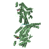
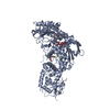
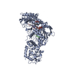
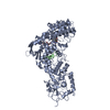
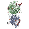
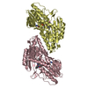
- PDB-1jmo: Crystal Structure of the Heparin Cofactor II-S195A Thrombin Complex -
+ Open data

- Basic information
| Entry | Database: PDB / ID: 1jmo | |||||||||
|---|---|---|---|---|---|---|---|---|---|---|
| Title | Crystal Structure of the Heparin Cofactor II-S195A Thrombin Complex | |||||||||
 Components Components |
| |||||||||
 Keywords Keywords | BLOOD CLOTTING / serpin / thrombin / protease / inhibition / inhibitor | |||||||||
| Function / homology |  Function and homology information Function and homology information: / thrombospondin receptor activity / thrombin / thrombin-activated receptor signaling pathway / Defective factor XII causes hereditary angioedema / negative regulation of astrocyte differentiation / endopeptidase inhibitor activity / regulation of blood coagulation / neutrophil-mediated killing of gram-negative bacterium / positive regulation of phospholipase C-activating G protein-coupled receptor signaling pathway ...: / thrombospondin receptor activity / thrombin / thrombin-activated receptor signaling pathway / Defective factor XII causes hereditary angioedema / negative regulation of astrocyte differentiation / endopeptidase inhibitor activity / regulation of blood coagulation / neutrophil-mediated killing of gram-negative bacterium / positive regulation of phospholipase C-activating G protein-coupled receptor signaling pathway / Defective F8 cleavage by thrombin / ligand-gated ion channel signaling pathway / Platelet Aggregation (Plug Formation) / positive regulation of collagen biosynthetic process / negative regulation of platelet activation / negative regulation of blood coagulation / negative regulation of fibrinolysis / blood coagulation, fibrin clot formation / positive regulation of blood coagulation / Transport of gamma-carboxylated protein precursors from the endoplasmic reticulum to the Golgi apparatus / : / Gamma-carboxylation of protein precursors / Removal of aminoterminal propeptides from gamma-carboxylated proteins / regulation of cytosolic calcium ion concentration / fibrinolysis / : / negative regulation of proteolysis / negative regulation of cytokine production involved in inflammatory response / Regulation of Complement cascade / acute-phase response / Cell surface interactions at the vascular wall / positive regulation of release of sequestered calcium ion into cytosol / Peptide ligand-binding receptors / growth factor activity / positive regulation of receptor signaling pathway via JAK-STAT / Post-translational protein phosphorylation / serine-type endopeptidase inhibitor activity / lipopolysaccharide binding / platelet activation / response to wounding / positive regulation of protein localization to nucleus / Golgi lumen / Regulation of Insulin-like Growth Factor (IGF) transport and uptake by Insulin-like Growth Factor Binding Proteins (IGFBPs) / positive regulation of reactive oxygen species metabolic process / chemotaxis / blood coagulation / positive regulation of insulin secretion / regulation of cell shape / antimicrobial humoral immune response mediated by antimicrobial peptide / heparin binding / Thrombin signalling through proteinase activated receptors (PARs) / positive regulation of cell growth / blood microparticle / G alpha (q) signalling events / cell surface receptor signaling pathway / positive regulation of phosphatidylinositol 3-kinase/protein kinase B signal transduction / endoplasmic reticulum lumen / receptor ligand activity / signaling receptor binding / serine-type endopeptidase activity / calcium ion binding / positive regulation of cell population proliferation / proteolysis / : / extracellular exosome / extracellular region / plasma membrane Similarity search - Function | |||||||||
| Biological species |  Homo sapiens (human) Homo sapiens (human) | |||||||||
| Method |  X-RAY DIFFRACTION / SYNCHROTRON / MOLECULAR REPLACEMENT / Resolution: 2.2 Å X-RAY DIFFRACTION / SYNCHROTRON / MOLECULAR REPLACEMENT / Resolution: 2.2 Å | |||||||||
 Authors Authors | Baglin, T.P. / Carrell, R.W. / Esmon, C.T. / Huntington, J.A. | |||||||||
 Citation Citation | Journal: Proc.Natl.Acad.Sci.USA / Year: 2002 Title: Crystal structures of native and thrombin-complexed heparin cofactor II reveal a multistep allosteric mechanism. Authors: Baglin, T.P. / Carrell, R.W. / Church, F.C. / Esmon, C.T. / Huntington, J.A. | |||||||||
| History |
|
- Structure visualization
| Structure viewer | Molecule: MolmilJmol/JSmol |
|---|
- Downloads & links
-Download
| PDBx/mmCIF format | 1jmo.cif.gz | 171.7 KB | Display | PDBx/mmCIF format |
|---|---|---|---|---|
| PDB format | pdb1jmo.ent.gz | 132.8 KB | Display | PDB format |
| PDBx/mmJSON format | 1jmo.json.gz | Tree view | PDBx/mmJSON format | |
| Others |  Other downloads Other downloads |
-Validation report
| Arichive directory | https://data.pdbj.org/pub/pdb/validation_reports/jm/1jmoftp://data.pdbj.org/pub/pdb/validation_reports/jm/1jmo | HTTPS FTP |
|---|
-Related structure data


-Links
 PDBj
PDBj


- Assembly
| Deposited unit | 
| ||||||||||
|---|---|---|---|---|---|---|---|---|---|---|---|
| 1 |
| ||||||||||
| Unit cell |
|
-Components
-Protein/peptide , 1 types, 1 molecules L
| #1: Protein/peptide | Mass: 5483.981 Da / Num. of mol.: 1 / Fragment: light chain Source method: isolated from a genetically manipulated source Details: contains extra 13 amino acids at the N-terminus / Source: (gene. exp.) Homo sapiens (human) / Cell line (production host): BHK culture / Organ (production host): kidney / Production host:   Cricetulus griseus (Chinese hamster) / References: UniProt: P00734 Cricetulus griseus (Chinese hamster) / References: UniProt: P00734 |
|---|
-Protein , 2 types, 2 molecules HA
| #2: Protein | Mass: 29921.414 Da / Num. of mol.: 1 / Fragment: heavy chain / Mutation: S195A Source method: isolated from a genetically manipulated source Source: (gene. exp.) Homo sapiens (human) / Cell line (production host): BHK culture / Organ (production host): kidney / Production host: Cricetulus griseus (Chinese hamster) / References: UniProt: P00734, thrombin |
|---|---|
| #3: Protein | Mass: 55186.961 Da / Num. of mol.: 1 / Source method: isolated from a natural source / Details: Plasma purified Human Heparin Cofactor II / Source: (natural) Homo sapiens (human) / Tissue fraction: blood plasma / References: UniProt: P05546 |
-Sugars , 2 types, 3 molecules 
| #4: Polysaccharide | 2-acetamido-2-deoxy-beta-D-glucopyranose-(1-4)-2-acetamido-2-deoxy-beta-D-glucopyranose Source method: isolated from a genetically manipulated source |
|---|---|
| #7: Sugar |  Type: D-saccharide, beta linking / Mass: 221.208 Da / Num. of mol.: 2 Type: D-saccharide, beta linking / Mass: 221.208 Da / Num. of mol.: 2Source method: isolated from a genetically manipulated source Formula: C8H15NO6 |
-Non-polymers , 3 types, 214 molecules 


| #5: Chemical | ChemComp-NA /  Mass: 22.990 Da / Num. of mol.: 1 / Source method: obtained synthetically / Formula: Na Mass: 22.990 Da / Num. of mol.: 1 / Source method: obtained synthetically / Formula: Na | ||
|---|---|---|---|
| #6: Chemical | ChemComp-MPD / (  Mass: 118.174 Da / Num. of mol.: 5 / Source method: obtained synthetically / Formula: C6H14O2 / Comment: precipitant*YM Mass: 118.174 Da / Num. of mol.: 5 / Source method: obtained synthetically / Formula: C6H14O2 / Comment: precipitant*YM#8: Water | ChemComp-HOH / | Mass: 18.015 Da / Num. of mol.: 208 / Source method: isolated from a natural source / Formula: H2O |
-Details
| Has protein modification | Y |
|---|
-Experimental details
-Experiment
| Experiment | Method: X-RAY DIFFRACTION / Number of used crystals: 1 |
|---|
- Sample preparation
| Crystal | Density Matthews: 4.68 Å3/Da / Density % sol: 73.75 % | ||||||||||||||||||||||||
|---|---|---|---|---|---|---|---|---|---|---|---|---|---|---|---|---|---|---|---|---|---|---|---|---|---|
| Crystal grow | Temperature: 298 K / Method: vapor diffusion, hanging drop / pH: 7.4 Details: NH4Cl, PEG 3350, pH 7.4, VAPOR DIFFUSION, HANGING DROP at 298K | ||||||||||||||||||||||||
| Crystal grow | *PLUS Method: unknown | ||||||||||||||||||||||||
| Components of the solutions | *PLUS
|
-Data collection
| Diffraction | Mean temperature: 100 K |
|---|---|
| Diffraction source | Source: SYNCHROTRON / Site: SRS  / Beamline: PX14.1 / Wavelength: 1.488 Å / Beamline: PX14.1 / Wavelength: 1.488 Å |
| Detector | Type: ADSC QUANTUM 4 / Detector: CCD / Date: Jan 30, 2001 / Details: mirrors |
| Radiation | Monochromator: liquid gallium / Protocol: SINGLE WAVELENGTH / Monochromatic (M) / Laue (L): M / Scattering type: x-ray |
| Radiation wavelength | Wavelength: 1.488 Å / Relative weight: 1 |
| Reflection | Resolution: 2.2→30 Å / Num. all: 84555 / Num. obs: 77077 / % possible obs: 91.2 % / Observed criterion σ(F): 2 / Observed criterion σ(I): 1 / Redundancy: 6.2 % / Biso Wilson estimate: 51.2 Å2 / Rmerge(I) obs: 0.086 / Rsym value: 0.081 / Net I/σ(I): 6.5 |
| Reflection shell | Resolution: 2.21→2.35 Å / Redundancy: 2.2 % / Rmerge(I) obs: 0.539 / Mean I/σ(I) obs: 1.5 / Num. unique all: 16159 / Rsym value: 0.434 / % possible all: 62.5 |
| Reflection | *PLUS Lowest resolution: 29.4 Å / Num. obs: 77117 / % possible obs: 91.1 % / Num. measured all: 475985 / Rmerge(I) obs: 0.08 |
- Processing
| Software |
| ||||||||||||||||||||||||||||
|---|---|---|---|---|---|---|---|---|---|---|---|---|---|---|---|---|---|---|---|---|---|---|---|---|---|---|---|---|---|
| Refinement | Method to determine structure: MOLECULAR REPLACEMENT Starting model: N-terminal half of native heparin cofactor II and native S195A thrombin Resolution: 2.2→29.44 Å / Isotropic thermal model: anisotropic / Cross valid method: THROUGHOUT / σ(F): 0 / σ(I): 0 / Stereochemistry target values: Engh & Huber / Details: maximum likelihood target of CNS
| ||||||||||||||||||||||||||||
| Displacement parameters | Biso mean: 55 Å2
| ||||||||||||||||||||||||||||
| Refine analyze |
| ||||||||||||||||||||||||||||
| Refinement step | Cycle: LAST / Resolution: 2.2→29.44 Å
| ||||||||||||||||||||||||||||
| Refine LS restraints |
| ||||||||||||||||||||||||||||
| LS refinement shell | Resolution: 2.2→2.34 Å / Rfactor Rfree error: 0.033
| ||||||||||||||||||||||||||||
| Refine LS restraints | *PLUS
| ||||||||||||||||||||||||||||
| LS refinement shell | *PLUS Rfactor Rfree: 0.34 |
