 Movie
Movie Controller

[English] 日本語
 Yorodumi
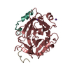
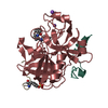
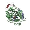
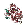
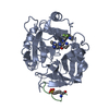
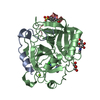
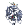
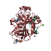
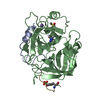
- PDB-1qbv: CRYSTAL STRUCTURE OF THROMBIN COMPLEXED WITH AN GUANIDINE-MIMETIC... -
+ Open data

- Basic information
| Entry | Database: PDB / ID: 1qbv | ||||||
|---|---|---|---|---|---|---|---|
| Title | CRYSTAL STRUCTURE OF THROMBIN COMPLEXED WITH AN GUANIDINE-MIMETIC INHIBITOR | ||||||
 Components Components |
| ||||||
 Keywords Keywords | HYDROLASE/HYDROLASE INHIBITOR / THROMBIN / INHIBITOR / 3DP / HYDROLASE / HYDROLASE-HYDROLASE INHIBITOR COMPLEX | ||||||
| Function / homology |  Function and homology information Function and homology information: / thrombospondin receptor activity / thrombin / thrombin-activated receptor signaling pathway / Defective factor XII causes hereditary angioedema / negative regulation of astrocyte differentiation / regulation of blood coagulation / neutrophil-mediated killing of gram-negative bacterium / positive regulation of phospholipase C-activating G protein-coupled receptor signaling pathway / Defective F8 cleavage by thrombin ...: / thrombospondin receptor activity / thrombin / thrombin-activated receptor signaling pathway / Defective factor XII causes hereditary angioedema / negative regulation of astrocyte differentiation / regulation of blood coagulation / neutrophil-mediated killing of gram-negative bacterium / positive regulation of phospholipase C-activating G protein-coupled receptor signaling pathway / Defective F8 cleavage by thrombin / ligand-gated ion channel signaling pathway / Platelet Aggregation (Plug Formation) / positive regulation of collagen biosynthetic process / negative regulation of platelet activation / negative regulation of blood coagulation / negative regulation of fibrinolysis / blood coagulation, fibrin clot formation / positive regulation of blood coagulation / Transport of gamma-carboxylated protein precursors from the endoplasmic reticulum to the Golgi apparatus / : / Gamma-carboxylation of protein precursors / Removal of aminoterminal propeptides from gamma-carboxylated proteins / regulation of cytosolic calcium ion concentration / fibrinolysis / : / negative regulation of proteolysis / negative regulation of cytokine production involved in inflammatory response / Regulation of Complement cascade / positive regulation of release of sequestered calcium ion into cytosol / acute-phase response / Cell surface interactions at the vascular wall / Peptide ligand-binding receptors / growth factor activity / positive regulation of receptor signaling pathway via JAK-STAT / serine-type endopeptidase inhibitor activity / lipopolysaccharide binding / platelet activation / positive regulation of protein localization to nucleus / response to wounding / Golgi lumen / Regulation of Insulin-like Growth Factor (IGF) transport and uptake by Insulin-like Growth Factor Binding Proteins (IGFBPs) / positive regulation of reactive oxygen species metabolic process / blood coagulation / positive regulation of insulin secretion / regulation of cell shape / antimicrobial humoral immune response mediated by antimicrobial peptide / heparin binding / Thrombin signalling through proteinase activated receptors (PARs) / positive regulation of cell growth / blood microparticle / G alpha (q) signalling events / positive regulation of phosphatidylinositol 3-kinase/protein kinase B signal transduction / cell surface receptor signaling pathway / endoplasmic reticulum lumen / receptor ligand activity / serine-type endopeptidase activity / signaling receptor binding / calcium ion binding / positive regulation of cell population proliferation / proteolysis / : / extracellular exosome / extracellular region / plasma membrane Similarity search - Function | ||||||
| Biological species |  Homo sapiens (human) Homo sapiens (human) Hirudo medicinalis (medicinal leech) Hirudo medicinalis (medicinal leech) | ||||||
| Method |  X-RAY DIFFRACTION / Resolution: 1.8 Å X-RAY DIFFRACTION / Resolution: 1.8 Å | ||||||
 Authors Authors | Bone, R. / Lu, T. / Illig, C.R. / Soll, R.M. / Spurlino, J.C. | ||||||
 Citation Citation | Journal: J.Med.Chem. / Year: 1998 Title: Structural analysis of thrombin complexed with potent inhibitors incorporating a phenyl group as a peptide mimetic and aminopyridines as guanidine substitutes. Authors: Bone, R. / Lu, T. / Illig, C.R. / Soll, R.M. / Spurlino, J.C. | ||||||
| History |
|
- Structure visualization
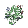
| Structure viewer | Molecule: MolmilJmol/JSmol |
|---|
- Downloads & links
-Download
| PDBx/mmCIF format | 1qbv.cif.gz | 76.4 KB | Display | PDBx/mmCIF format |
|---|---|---|---|---|
| PDB format | pdb1qbv.ent.gz | 55.4 KB | Display | PDB format |
| PDBx/mmJSON format | 1qbv.json.gz | Tree view | PDBx/mmJSON format | |
| Others |  Other downloads Other downloads |
-Validation report
| Arichive directory | https://data.pdbj.org/pub/pdb/validation_reports/qb/1qbvftp://data.pdbj.org/pub/pdb/validation_reports/qb/1qbv | HTTPS FTP |
|---|
-Related structure data
| Similar structure data |
|---|


-Links
 PDBj
PDBj


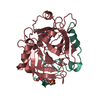
- Assembly
| Deposited unit | 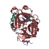
| ||||||||
|---|---|---|---|---|---|---|---|---|---|
| 1 |
| ||||||||
| Unit cell |
|
-Components
| #1: Protein/peptide | Mass: 4096.534 Da / Num. of mol.: 1 / Fragment: LIGHT CHAIN Source method: isolated from a genetically manipulated source Source: (gene. exp.) Homo sapiens (human) / Description: Homo sapiens / References: UniProt: P00734, thrombin |
|---|---|
| #2: Protein | Mass: 29780.219 Da / Num. of mol.: 1 / Fragment: HEAVY CHAIN Source method: isolated from a genetically manipulated source Source: (gene. exp.) Homo sapiens (human) / Description: Homo sapiens / References: UniProt: P00734, thrombin |
| #3: Protein/peptide | Mass: 1491.528 Da / Num. of mol.: 1 / Fragment: residues 55-65 Source method: isolated from a genetically manipulated source Source: (gene. exp.) Hirudo medicinalis (medicinal leech) / Genus: Hirudinaria / References: UniProt: P28504 |
| #4: Chemical | ChemComp-PPX / [  Type: peptide-like / Mass: 381.471 Da / Num. of mol.: 1 / Source method: obtained synthetically / Formula: C21H27N5O2 Type: peptide-like / Mass: 381.471 Da / Num. of mol.: 1 / Source method: obtained synthetically / Formula: C21H27N5O2 |
| #5: Water | ChemComp-HOH /  Mass: 18.015 Da / Num. of mol.: 121 / Source method: isolated from a natural source / Formula: H2O Mass: 18.015 Da / Num. of mol.: 121 / Source method: isolated from a natural source / Formula: H2O |
| Has protein modification | Y |
-Experimental details
-Experiment
| Experiment | Method: X-RAY DIFFRACTION / Number of used crystals: 1 |
|---|
- Sample preparation
| Crystal | Density Matthews: 2.57 Å3/Da / Density % sol: 52.15 % |
|---|---|
| Crystal grow | Temperature: 277.15 K / Method: vapor diffusion, hanging drop / pH: 7.3 Details: PEG 8000, phosphate, NaCl, NAN3, pH 7.3, VAPOR DIFFUSION, HANGING DROP, temperature 277.15K |
| Crystal grow | *PLUS Details: Skrzypczak-Jankun, E., (1991) J. Mol. Biol., 221, 1379. |
-Data collection
| Diffraction | Mean temperature: 288 K |
|---|---|
| Diffraction source | Source: ROTATING ANODE / Type: ENRAF-NONIUS FR571 / Wavelength: 1.5418 |
| Detector | Type: RIGAKU RAXIS II / Detector: IMAGE PLATE / Date: May 1, 1995 |
| Radiation | Protocol: SINGLE WAVELENGTH / Monochromatic (M) / Laue (L): M / Scattering type: x-ray |
| Radiation wavelength | Wavelength: 1.5418 Å / Relative weight: 1 |
| Reflection | Resolution: 1.8→8 Å / Num. all: 28984 / Num. obs: 26244 / % possible obs: 85.8 % / Observed criterion σ(F): 2 / Observed criterion σ(I): 2 / Redundancy: 5.2 % / Biso Wilson estimate: 12.4 Å2 / Rmerge(I) obs: 0.054 / Net I/σ(I): 12.2 |
| Reflection shell | Resolution: 1.8→1.88 Å / Redundancy: 2.4 % / Rmerge(I) obs: 0.091 / Num. unique all: 2008 / % possible all: 60.4 |
| Reflection | *PLUS Rmerge(I) obs: 0.083 |
- Processing
| Software |
| ||||||||||||||||
|---|---|---|---|---|---|---|---|---|---|---|---|---|---|---|---|---|---|
| Refinement | Resolution: 1.8→8 Å / Data cutoff high absF: 10000000 / Data cutoff low absF: 0 / Cross valid method: THROUGHOUT / σ(F): 2 / σ(I): 2 / Stereochemistry target values: Engh & Huber
| ||||||||||||||||
| Displacement parameters | Biso mean: 21.6 Å2 | ||||||||||||||||
| Refine analyze | Luzzati coordinate error obs: 0.26 Å / Luzzati d res low obs: 5 Å / Luzzati sigma a obs: 0.22 Å | ||||||||||||||||
| Refinement step | Cycle: LAST / Resolution: 1.8→8 Å
| ||||||||||||||||
| Refine LS restraints |
| ||||||||||||||||
| LS refinement shell | Resolution: 1.8→1.86 Å / Total num. of bins used: 10
| ||||||||||||||||
| Software | *PLUS Name: X-PLOR(ONLINE) / Version: 98 / Classification: refinement | ||||||||||||||||
| Refinement | *PLUS σ(F): 2 | ||||||||||||||||
| Solvent computation | *PLUS | ||||||||||||||||
| Displacement parameters | *PLUS Biso mean: 21.6 Å2 | ||||||||||||||||
| LS refinement shell | *PLUS Rfactor Rwork: 0.285 |
