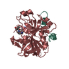
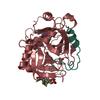
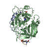
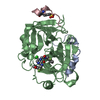
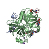
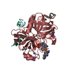
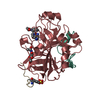
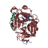
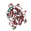
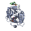
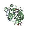
Entry Database : PDB / ID : 2a2xTitle Orally Active Thrombin Inhibitors in Complex with Thrombin Inh12 Thrombin heavy chain Thrombin light chain synthetic peptide Keywords / / / Function / homology Function Domain/homology Component
/ / / / / / / / / / / / / / / / / / / / / / / / / / / / / / / / / / / / / / / / / / / / / / / / / / / / / / / / / / / / / / / / / / / / / / / / / / / / / / / / / / / / / / / / / / / / / / / / / / / / / / / / / / / / / / / / / Biological species Homo sapiens (human)Synthetic (others) Method / / Resolution : 2.44 Å Authors Lange, U.E.W. / Baucke, D. / Hornberger, W. / Mack, H. / Seitz, W. / Hoeffken, H.W. Journal : BIOORG.MED.CHEM.LETT. / Year : 2006Title : Orally active thrombin inhibitors. Part 2: optimization of the P2-moietyAuthors : Lange, U.E.W. / Baucke, D. / Hornberger, W. / Mack, H. / Seitz, W. / Hoeffken, H.W. History Deposition Jun 23, 2005 Deposition site / Processing site Revision 1.0 Nov 14, 2006 Provider / Type Revision 1.1 Apr 30, 2008 Group Revision 1.2 Jul 13, 2011 Group Atomic model / Database references ... Atomic model / Database references / Derived calculations / Non-polymer description / Structure summary / Version format compliance Revision 1.3 Dec 12, 2012 Group Revision 1.4 Oct 11, 2017 Group / Category / Item Revision 1.5 Apr 4, 2018 Group / Category / Item Revision 1.6 Aug 23, 2023 Group Data collection / Database references ... Data collection / Database references / Derived calculations / Refinement description / Structure summary Category chem_comp / chem_comp_atom ... chem_comp / chem_comp_atom / chem_comp_bond / database_2 / pdbx_initial_refinement_model / struct_conn / struct_site Item _chem_comp.pdbx_synonyms / _database_2.pdbx_DOI ... _chem_comp.pdbx_synonyms / _database_2.pdbx_DOI / _database_2.pdbx_database_accession / _struct_conn.pdbx_leaving_atom_flag / _struct_site.pdbx_auth_asym_id / _struct_site.pdbx_auth_comp_id / _struct_site.pdbx_auth_seq_id
Show all Show less
 Movie
Movie Controller


 Open data
 Basic information
 Components
 Keywords
 Function and homology information
 Homo sapiens (human)
 X-RAY DIFFRACTION /
  Authors
 Citation
 Structure visualization
 Downloads & links
 Other downloads
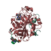

 PDBj
PDBj


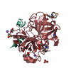
 Assembly
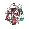


 Type: Oligopeptide / Class: Anticoagulant, Antithrombotic / Mass: 1514.605 Da / Num. of mol.: 1 / Source method: obtained synthetically / Details: Synthetic peptide / Source: (synth.) Synthetic (others) / References: HIRUDIN ANALOGUE
 Mass: 446.543 Da / Num. of mol.: 1 / Source method: obtained synthetically / Formula: C22H34N6O4
 Mass: 18.015 Da / Num. of mol.: 174 / Source method: isolated from a natural source / Formula: H2O
 Sample preparation
 Processing