 Movie
Movie Controller

[English] 日本語
 Yorodumi
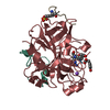
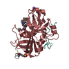
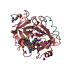
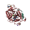
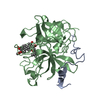
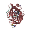
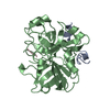
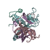
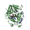
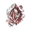
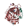
- PDB-1e0f: Crystal structure of the human alpha-thrombin-haemadin complex: a... -
+ Open data

- Basic information
| Entry | Database: PDB / ID: 1e0f | ||||||
|---|---|---|---|---|---|---|---|
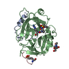
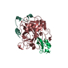
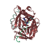
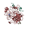
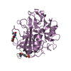
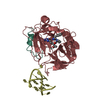
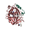
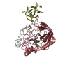
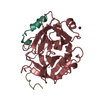
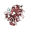
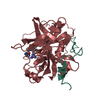
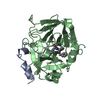
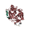
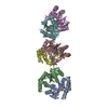
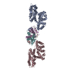
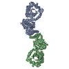
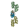
| Title | Crystal structure of the human alpha-thrombin-haemadin complex: an exosite II-binding inhibitor | ||||||
 Components Components |
| ||||||
 Keywords Keywords | HYDROLASE / COAGULATION/CRYSTAL STRUCTURE/HEPARIN-B / COAGULATION/CRYSTAL STRUCTURE/HEPARIN-BINDING SITE/ HIRUDIN/THROMBIN INHIBITOR | ||||||
| Function / homology |  Function and homology information Function and homology information: / thrombospondin receptor activity / thrombin / thrombin-activated receptor signaling pathway / Defective factor XII causes hereditary angioedema / negative regulation of astrocyte differentiation / regulation of blood coagulation / neutrophil-mediated killing of gram-negative bacterium / positive regulation of phospholipase C-activating G protein-coupled receptor signaling pathway / Defective F8 cleavage by thrombin ...: / thrombospondin receptor activity / thrombin / thrombin-activated receptor signaling pathway / Defective factor XII causes hereditary angioedema / negative regulation of astrocyte differentiation / regulation of blood coagulation / neutrophil-mediated killing of gram-negative bacterium / positive regulation of phospholipase C-activating G protein-coupled receptor signaling pathway / Defective F8 cleavage by thrombin / ligand-gated ion channel signaling pathway / Platelet Aggregation (Plug Formation) / positive regulation of collagen biosynthetic process / negative regulation of platelet activation / negative regulation of blood coagulation / negative regulation of fibrinolysis / blood coagulation, fibrin clot formation / positive regulation of blood coagulation / Transport of gamma-carboxylated protein precursors from the endoplasmic reticulum to the Golgi apparatus / : / Gamma-carboxylation of protein precursors / Removal of aminoterminal propeptides from gamma-carboxylated proteins / regulation of cytosolic calcium ion concentration / fibrinolysis / : / negative regulation of proteolysis / negative regulation of cytokine production involved in inflammatory response / Regulation of Complement cascade / acute-phase response / Cell surface interactions at the vascular wall / positive regulation of release of sequestered calcium ion into cytosol / Peptide ligand-binding receptors / growth factor activity / positive regulation of receptor signaling pathway via JAK-STAT / serine-type endopeptidase inhibitor activity / lipopolysaccharide binding / platelet activation / response to wounding / positive regulation of protein localization to nucleus / Golgi lumen / Regulation of Insulin-like Growth Factor (IGF) transport and uptake by Insulin-like Growth Factor Binding Proteins (IGFBPs) / positive regulation of reactive oxygen species metabolic process / blood coagulation / positive regulation of insulin secretion / regulation of cell shape / antimicrobial humoral immune response mediated by antimicrobial peptide / heparin binding / Thrombin signalling through proteinase activated receptors (PARs) / positive regulation of cell growth / blood microparticle / G alpha (q) signalling events / cell surface receptor signaling pathway / positive regulation of phosphatidylinositol 3-kinase/protein kinase B signal transduction / endoplasmic reticulum lumen / receptor ligand activity / signaling receptor binding / serine-type endopeptidase activity / calcium ion binding / positive regulation of cell population proliferation / proteolysis / : / extracellular exosome / extracellular region / plasma membrane Similarity search - Function | ||||||
| Biological species |  HAEMADIPSA SYLVESTRIS (Indian leech) HAEMADIPSA SYLVESTRIS (Indian leech) HOMO SAPIENS (human) HOMO SAPIENS (human) | ||||||
| Method |  X-RAY DIFFRACTION / MOLECULAR REPLACEMENT / Resolution: 3.1 Å X-RAY DIFFRACTION / MOLECULAR REPLACEMENT / Resolution: 3.1 Å | ||||||
 Authors Authors | Richardson, J.L. / Kroeger, B. / Hoefken, W. / Pereira, P. / Huber, R. / Bode, W. / Fuentes-Prior, P. | ||||||
 Citation Citation | Journal: Embo J. / Year: 2000 Title: Crystal Structure of the Human Alpha-Thrombin-Haemadin Complex: An Exosite II-Binding Inhibitor Authors: Richardson, J.L. / Kroeger, B. / Hoeffken, W. / Sadler, J.E. / Pereira, P. / Huber, R. / Bode, W. / Fuentes-Prior, P. #1: Journal: J.Biol.Chem. / Year: 1993 Title: Isolation, Sequence Analysis, and Cloning of Haemadin an Anticoagulant Peptide from the Indian Leech Authors: Strube, K.-H. / Kroeger, B. / Bialojan, S. / Otte, M. / Dodt, J. #2: Journal: Protein Sci. / Year: 1992Title: The Refined 1.9 Angstrom X-Ray Crystal Structure of D-Phe-Pro-Arg-Chloromethylketone Inhibited Human Alpha Thrombin: Structure Analysis, Overall Structure, Electrostatic Properties, Detailed ...Title: The Refined 1.9 Angstrom X-Ray Crystal Structure of D-Phe-Pro-Arg-Chloromethylketone Inhibited Human Alpha Thrombin: Structure Analysis, Overall Structure, Electrostatic Properties, Detailed Active Site Geometry and Structure Function Relatioships Authors: Bode, W. / Turk, D. / Karshikov, A. | ||||||
| History |
| ||||||
| Remark 700 | SHEET DETERMINATION METHOD: DSSP THE SHEETS PRESENTED AS "B" IN EACH CHAIN ON SHEET RECORDS BELOW ... SHEET DETERMINATION METHOD: DSSP THE SHEETS PRESENTED AS "B" IN EACH CHAIN ON SHEET RECORDS BELOW IS ACTUALLY AN 6-STRANDED BARREL THIS IS REPRESENTED BY A 7-STRANDED SHEET IN WHICH THE FIRST AND LAST STRANDS ARE IDENTICAL. |
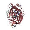
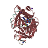
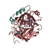
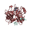
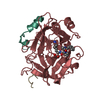
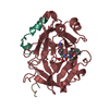
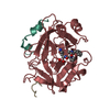
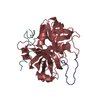
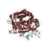
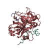
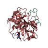
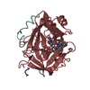
- Structure visualization
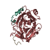
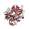
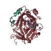
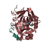
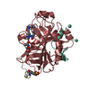
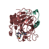
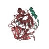
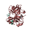
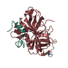
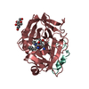
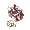
| Structure viewer | Molecule: MolmilJmol/JSmol |
|---|
- Downloads & links
-Download
| PDBx/mmCIF format | 1e0f.cif.gz | 217.9 KB | Display | PDBx/mmCIF format |
|---|---|---|---|---|
| PDB format | pdb1e0f.ent.gz | 174.9 KB | Display | PDB format |
| PDBx/mmJSON format | 1e0f.json.gz | Tree view | PDBx/mmJSON format | |
| Others |  Other downloads Other downloads |
-Validation report
| Arichive directory | https://data.pdbj.org/pub/pdb/validation_reports/e0/1e0fftp://data.pdbj.org/pub/pdb/validation_reports/e0/1e0f | HTTPS FTP |
|---|
-Related structure data
| Related structure data | 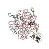 4htcS S: Starting model for refinement |
|---|---|
| Similar structure data |


-Links
 PDBj
PDBj


- Assembly
| Deposited unit | 
| ||||||||
|---|---|---|---|---|---|---|---|---|---|
| 1 |
| ||||||||
| Unit cell |
|
-Components
| #1: Protein/peptide | Mass: 4096.534 Da / Num. of mol.: 3 / Source method: isolated from a natural source Details: HUMAN THROMBIN WAS PURIFIED FROM HUMAN SERUM ACCORDING TO REPORTED PROTOCOLS Source: (natural) HOMO SAPIENS (human) / Tissue: BLOOD / References: UniProt: P00734#2: Protein | Mass: 29792.273 Da / Num. of mol.: 3 / Fragment: NO / Source method: isolated from a natural source Details: HUMAN THROMBIN WAS PURIFIED FROM HUMAN SERUM ACCORDING TO REPORTED PROTOCOLS Source: (natural) HOMO SAPIENS (human) / Tissue: BLOOD / References: UniProt: P00734, thrombin#3: Protein | Mass: 6262.853 Da / Num. of mol.: 3 / Fragment: NO Source method: isolated from a genetically manipulated source Source: (gene. exp.) HAEMADIPSA SYLVESTRIS (Indian leech)Description: RECOMBINANTLY EXPRESSED IN E. COLI AS A MALTOSE BINDING PROTEIN CONJUGATE Plasmid: PMAL-P2 / Production host:  #4: Water | ChemComp-HOH / |  Mass: 18.015 Da / Num. of mol.: 67 / Source method: isolated from a natural source / Formula: H2O Mass: 18.015 Da / Num. of mol.: 67 / Source method: isolated from a natural source / Formula: H2OHas protein modification | Y | Sequence details | CHYMOTRYPSIN NUMBERING (RATHER THAN SEQUENTIAL) SYSTEM IS USED, BASED ON THE TOPOLOGICAL ALIGNMENT ...CHYMOTRYPS | |
|---|
-Experimental details
-Experiment
| Experiment | Method: X-RAY DIFFRACTION / Number of used crystals: 2 |
|---|
- Sample preparation
| Crystal | Density Matthews: 3.08 Å3/Da / Density % sol: 60 % | ||||||||||||||||||||||||||||||||||||||||||||||||||||||||
|---|---|---|---|---|---|---|---|---|---|---|---|---|---|---|---|---|---|---|---|---|---|---|---|---|---|---|---|---|---|---|---|---|---|---|---|---|---|---|---|---|---|---|---|---|---|---|---|---|---|---|---|---|---|---|---|---|---|
| Crystal grow | Method: vapor diffusion, sitting drop / pH: 5.56 Details: VAPOUR-DIFFUSION SITTING DROP,0.1 M NA CITRATE PH 5.56 14% (W/V) PEG4000, 12.5% (V/V) ISOPROPANOL | ||||||||||||||||||||||||||||||||||||||||||||||||||||||||
| Crystal grow | *PLUS Temperature: 22 ℃ / Method: vapor diffusionDetails: drop consists of equal amounts of protein and precipitant solutions | ||||||||||||||||||||||||||||||||||||||||||||||||||||||||
| Components of the solutions | *PLUS
|
-Data collection
| Diffraction | Mean temperature: 289 K |
|---|---|
| Diffraction source | Source: ROTATING ANODE / Type: RIGAKU RUH2R / Wavelength: 1.5418 |
| Detector | Type: MARRESEARCH / Detector: IMAGE PLATE / Date: Sep 15, 1998 |
| Radiation | Monochromator: NI FILTER / Protocol: SINGLE WAVELENGTH / Monochromatic (M) / Laue (L): M / Scattering type: x-ray |
| Radiation wavelength | Wavelength: 1.5418 Å / Relative weight: 1 |
| Reflection | Resolution: 3.1→32.791 Å / Num. obs: 23938 / Rmerge(I) obs: 0.109 |
| Reflection | *PLUS % possible obs: 81.2 % / Num. measured all: 126754 |
- Processing
| Software |
| ||||||||||||||||||||||||||||||||||||||||||||||||||||||||||||
|---|---|---|---|---|---|---|---|---|---|---|---|---|---|---|---|---|---|---|---|---|---|---|---|---|---|---|---|---|---|---|---|---|---|---|---|---|---|---|---|---|---|---|---|---|---|---|---|---|---|---|---|---|---|---|---|---|---|---|---|---|---|
| Refinement | Method to determine structure: MOLECULAR REPLACEMENT Starting model: 4HTC Resolution: 3.1→10 Å / Cross valid method: THROUGHOUT / σ(F): 0
| ||||||||||||||||||||||||||||||||||||||||||||||||||||||||||||
| Refinement step | Cycle: LAST / Resolution: 3.1→10 Å
| ||||||||||||||||||||||||||||||||||||||||||||||||||||||||||||
| Refine LS restraints |
| ||||||||||||||||||||||||||||||||||||||||||||||||||||||||||||
| Software | *PLUS Name: X-PLOR / Version: 3.851 / Classification: refinement | ||||||||||||||||||||||||||||||||||||||||||||||||||||||||||||
| Refine LS restraints | *PLUS
|
