 Movie
Movie Controller


+ Open data

- Basic information
| Entry | Database: PDB / ID: 2i3s | ||||||
|---|---|---|---|---|---|---|---|
| Title | Bub3 complex with Bub1 GLEBS motif | ||||||
 Components Components |
| ||||||
 Keywords Keywords | CELL CYCLE / WD40 protein / beta-propeller / GLEBS motif / mitotic spindle checkpoint | ||||||
| Function / homology |  Function and homology information Function and homology informationbub1-bub3 complex / Inactivation of APC/C via direct inhibition of the APC/C complex / mitotic DNA integrity checkpoint signaling / mitotic checkpoint complex / sister chromatid biorientation / meiotic sister chromatid cohesion, centromeric / outer kinetochore / condensed chromosome, centromeric region / protein localization to kinetochore / positive regulation of protein autoubiquitination ...bub1-bub3 complex / Inactivation of APC/C via direct inhibition of the APC/C complex / mitotic DNA integrity checkpoint signaling / mitotic checkpoint complex / sister chromatid biorientation / meiotic sister chromatid cohesion, centromeric / outer kinetochore / condensed chromosome, centromeric region / protein localization to kinetochore / positive regulation of protein autoubiquitination / mitotic spindle assembly checkpoint signaling / macroautophagy / ubiquitin binding / kinetochore / protein kinase activity / non-specific serine/threonine protein kinase / protein serine kinase activity / protein serine/threonine kinase activity / nucleoplasm / ATP binding / identical protein binding / nucleus Similarity search - Function | ||||||
| Biological species |  | ||||||
| Method |  X-RAY DIFFRACTION / SYNCHROTRON / MOLECULAR REPLACEMENT / Resolution: 1.9 Å X-RAY DIFFRACTION / SYNCHROTRON / MOLECULAR REPLACEMENT / Resolution: 1.9 Å | ||||||
 Authors Authors | Larsen, N.A. / Harrison, S.C. | ||||||
 Citation Citation | Journal: Proc.Natl.Acad.Sci.Usa / Year: 2007 Title: Structural analysis of Bub3 interactions in the mitotic spindle checkpoint. Authors: Larsen, N.A. / Al-Bassam, J. / Wei, R.R. / Harrison, S.C. #1: Journal: J.Mol.Biol. / Year: 2004Title: Crystal structure of the spindle assembly checkpoint protein Bub3. Authors: Larsen, N.A. / Harrison, S.C. | ||||||
| History |
|
- Structure visualization
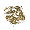
| Structure viewer | Molecule: MolmilJmol/JSmol |
|---|
- Downloads & links
-Download
| PDBx/mmCIF format | 2i3s.cif.gz | 240 KB | Display | PDBx/mmCIF format |
|---|---|---|---|---|
| PDB format | pdb2i3s.ent.gz | 191.9 KB | Display | PDB format |
| PDBx/mmJSON format | 2i3s.json.gz | Tree view | PDBx/mmJSON format | |
| Others |  Other downloads Other downloads |
-Validation report
| Arichive directory | https://data.pdbj.org/pub/pdb/validation_reports/i3/2i3sftp://data.pdbj.org/pub/pdb/validation_reports/i3/2i3s | HTTPS FTP |
|---|
-Related structure data
| Related structure data | 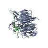 2i3tC 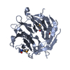 1u4cS S: Starting model for refinement C: citing same article ( |
|---|---|
| Similar structure data |


-Links
 PDBj
PDBj- Assembly
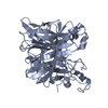
| Deposited unit | 
| ||||||||
|---|---|---|---|---|---|---|---|---|---|
| 1 | 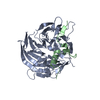
| ||||||||
| 2 | 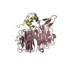
| ||||||||
| 3 | 
| ||||||||
| Unit cell |
| ||||||||
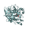
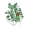
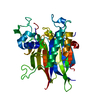
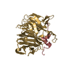
| Details | The biological assembly consists of a single heterodimer of Bub3 and Bub1 GLEBS peptide. There are 3 heterodimers in the asymmetric unit. |
-Components
| #1: Protein | Mass: 39554.633 Da / Num. of mol.: 3 Source method: isolated from a genetically manipulated source Source: (gene. exp.) Gene: BUB3, YOR026W, OR26.16 / Production host:  #2: Protein/peptide | Mass: 4349.952 Da / Num. of mol.: 3 / Source method: obtained synthetically / Details: Chemically synthesized peptide. / Source: (synth.) References: UniProt: P41695, non-specific serine/threonine protein kinase #3: Water | ChemComp-HOH / |  Mass: 18.015 Da / Num. of mol.: 644 / Source method: isolated from a natural source / Formula: H2O Mass: 18.015 Da / Num. of mol.: 644 / Source method: isolated from a natural source / Formula: H2O |
|---|
-Experimental details
-Experiment
| Experiment | Method: X-RAY DIFFRACTION / Number of used crystals: 1 |
|---|
- Sample preparation
| Crystal | Density Matthews: 2.15 Å3/Da / Density % sol: 42.72 % |
|---|---|
| Crystal grow | Temperature: 296 K / Method: vapor diffusion, hanging drop / pH: 6.4 Details: 18% PEG 3K 200 mM NaCl 100 mM Cacodylate, pH 6.4, VAPOR DIFFUSION, HANGING DROP, temperature 296K |
-Data collection
| Diffraction | Mean temperature: 120 K |
|---|---|
| Diffraction source | Source: SYNCHROTRON / Site: ALS  / Beamline: 8.2.2 / Wavelength: 0.9778 Å / Beamline: 8.2.2 / Wavelength: 0.9778 Å |
| Detector | Type: ADSC QUANTUM 315 / Detector: CCD / Date: Aug 15, 2005 |
| Radiation | Protocol: SINGLE WAVELENGTH / Monochromatic (M) / Laue (L): M / Scattering type: x-ray |
| Radiation wavelength | Wavelength: 0.9778 Å / Relative weight: 1 |
| Reflection | Resolution: 1.9→50 Å / Num. obs: 79711 / % possible obs: 90.6 % / Observed criterion σ(F): 2.3 / Observed criterion σ(I): 2.3 / Rsym value: 0.054 |
| Reflection shell | Resolution: 1.9→1.97 Å / % possible all: 68.9 |
- Processing
| Software |
| ||||||||||||||||||||
|---|---|---|---|---|---|---|---|---|---|---|---|---|---|---|---|---|---|---|---|---|---|
| Refinement | Method to determine structure: MOLECULAR REPLACEMENT Starting model: PDB entry 1U4C Resolution: 1.9→50 Å / σ(F): 0 / Stereochemistry target values: Engh & Huber
| ||||||||||||||||||||
| Refine analyze | Luzzati coordinate error obs: 0.32 Å / Luzzati d res low obs: 5 Å / Luzzati sigma a obs: 0.29 Å | ||||||||||||||||||||
| Refinement step | Cycle: LAST / Resolution: 1.9→50 Å
| ||||||||||||||||||||
| Refine LS restraints |
|
