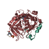
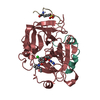
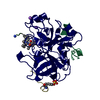
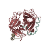
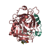
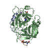
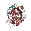
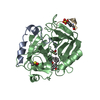
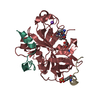
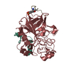
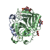
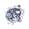
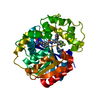
Entry Database : PDB / ID : 6eo9Title Crystal structure of thrombin in complex with a novel glucose-conjugated potent inhibitor (Prothrombin) x 2 Hirudin variant-2 Keywords / Function / homology Function Domain/homology Component
/ / / / / / / / / / / / / / / / / / / / / / / / / / / / / / / / / / / / / / / / / / / / / / / / / / / / / / / / / / / / / / / / / / / / / / / / / / / / / / / / / / / / / / / / / / / / / / / / / / / / / / / / / / / / / / / / / / / / / / Biological species Homo sapiens (human)Hirudo medicinalis (medicinal leech)Method / / / Resolution : 1.84 Å Authors Belviso, B.D. / Caliandro, R. / Aresta, B.M. / De Candia, M. / Altomare, C.D. Journal : J. Med. Chem. / Year : 2014Title: How a beta-D-glucoside side chain enhances binding affinity to thrombin of inhibitors bearing 2-chlorothiophene as P1 moiety: crystallography, fragment deconstruction study, and evaluation of ... Title : How a beta-D-glucoside side chain enhances binding affinity to thrombin of inhibitors bearing 2-chlorothiophene as P1 moiety: crystallography, fragment deconstruction study, and evaluation of antithrombotic properties.Authors : Belviso, B.D. / Caliandro, R. / de Candia, M. / Zaetta, G. / Lopopolo, G. / Incampo, F. / Colucci, M. / Altomare, C.D. History Deposition Oct 9, 2017 Deposition site / Processing site Supersession Dec 13, 2017 ID 4NZE Revision 1.0 Dec 13, 2017 Provider / Type Revision 1.1 Mar 7, 2018 Group / Category Item / _entity_src_gen.pdbx_host_org_scientific_nameRevision 1.2 Oct 16, 2019 Group / Category Revision 1.3 Nov 20, 2024 Group / Database references / Structure summaryCategory chem_comp_atom / chem_comp_bond ... chem_comp_atom / chem_comp_bond / database_2 / pdbx_entry_details / pdbx_modification_feature Item / _database_2.pdbx_database_accession
Show all Show less
 Movie
Movie Controller

 Yorodumi
 Open data
 Basic information
 Components
 Keywords
 Function and homology information
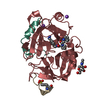
 Homo sapiens (human)
 Hirudo medicinalis (medicinal leech)
 X-RAY DIFFRACTION /
  Authors
 Citation
 Structure visualization
 Downloads & links
 Other downloads
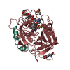


 PDBj
PDBj


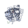
 Assembly


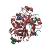
 Mass: 78.133 Da / Num. of mol.: 6 / Source method: obtained synthetically / Formula: C2H6OS / Comment: DMSO, precipitant*YM
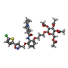
 Mass: 864.355 Da / Num. of mol.: 1 / Source method: obtained synthetically / Formula: C40H50ClN3O14S / Feature type: SUBJECT OF INVESTIGATION
 Sample preparation
 / Beamline: I24 / Wavelength: 0.9795 Å
 Processing