 Movie
Movie Controller

[English] 日本語
 Yorodumi
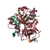
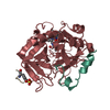
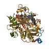
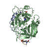
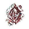
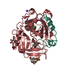
- PDB-3fbw: Structure of Rhodococcus rhodochrous haloalkane dehalogenase DhaA... -
+ Open data

- Basic information
| Entry | Database: PDB / ID: 3fbw | ||||||
|---|---|---|---|---|---|---|---|
| Title | Structure of Rhodococcus rhodochrous haloalkane dehalogenase DhaA mutant C176Y | ||||||
 Components Components | Haloalkane dehalogenase | ||||||
 Keywords Keywords | HYDROLASE / Detoxification / alpha/beta-Hydrolase / catalytic triad / halide-binding site | ||||||
| Function / homology |  Function and homology information Function and homology informationhaloalkane dehalogenase / haloalkane dehalogenase activity / response to toxic substance / membrane Similarity search - Function | ||||||
| Biological species |  Rhodococcus rhodochrous (bacteria) Rhodococcus rhodochrous (bacteria) | ||||||
| Method |  X-RAY DIFFRACTION / SYNCHROTRON / MOLECULAR REPLACEMENT / Resolution: 1.23 Å X-RAY DIFFRACTION / SYNCHROTRON / MOLECULAR REPLACEMENT / Resolution: 1.23 Å | ||||||
 Authors Authors | Dohnalek, J. / Stsiapanava, A. / Gavira, J.A. / Kuta Smatanova, I. / Kuty, M. | ||||||
 Citation Citation | Journal: Acta Crystallogr.,Sect.D / Year: 2010 Title: Atomic resolution studies of haloalkane dehalogenases DhaA04, DhaA14 and DhaA15 with engineered access tunnels. Authors: Stsiapanava, A. / Dohnalek, J. / Gavira, J.A. / Kuty, M. / Koudelakova, T. / Damborsky, J. / Kuta Smatanova, I. | ||||||
| History |
|
- Structure visualization
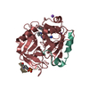
| Structure viewer | Molecule: MolmilJmol/JSmol |
|---|
- Downloads & links
-Download
| PDBx/mmCIF format | 3fbw.cif.gz | 157.7 KB | Display | PDBx/mmCIF format |
|---|---|---|---|---|
| PDB format | pdb3fbw.ent.gz | 121.9 KB | Display | PDB format |
| PDBx/mmJSON format | 3fbw.json.gz | Tree view | PDBx/mmJSON format | |
| Others |  Other downloads Other downloads |
-Validation report
| Arichive directory | https://data.pdbj.org/pub/pdb/validation_reports/fb/3fbwftp://data.pdbj.org/pub/pdb/validation_reports/fb/3fbw | HTTPS FTP |
|---|
-Related structure data
| Related structure data | 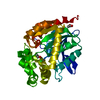 3fwhC 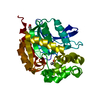 3g9xC 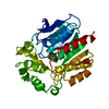 1bn6S C: citing same article ( S: Starting model for refinement |
|---|---|
| Similar structure data |

-Links
 PDBj
PDBj


- Assembly
| Deposited unit | 
| ||||||||
|---|---|---|---|---|---|---|---|---|---|
| 1 |
| ||||||||
| Unit cell |
|
-Components
| #1: Protein | Mass: 34170.898 Da / Num. of mol.: 1 / Mutation: C176Y Source method: isolated from a genetically manipulated source Details: The active site partially occupied by a small ligand and by a chloride ion Source: (gene. exp.) Rhodococcus rhodochrous (bacteria) / Strain: NCIMB 13064 / Gene: dhaA / Plasmid: pAQN / Production host: | ||||
|---|---|---|---|---|---|
| #2: Chemical | ChemComp-BEZ /   Mass: 122.121 Da / Num. of mol.: 1 / Source method: obtained synthetically / Formula: C7H6O2 Mass: 122.121 Da / Num. of mol.: 1 / Source method: obtained synthetically / Formula: C7H6O2 | ||||
| #3: Chemical | ChemComp-CL /   Mass: 35.453 Da / Num. of mol.: 6 / Source method: obtained synthetically / Formula: Cl Mass: 35.453 Da / Num. of mol.: 6 / Source method: obtained synthetically / Formula: Cl#4: Chemical |   Mass: 24.305 Da / Num. of mol.: 2 / Source method: obtained synthetically / Formula: Mg Mass: 24.305 Da / Num. of mol.: 2 / Source method: obtained synthetically / Formula: Mg#5: Water | ChemComp-HOH / |  Mass: 18.015 Da / Num. of mol.: 454 / Source method: isolated from a natural source / Formula: H2O Mass: 18.015 Da / Num. of mol.: 454 / Source method: isolated from a natural source / Formula: H2O |
-Experimental details
-Experiment
| Experiment | Method: X-RAY DIFFRACTION / Number of used crystals: 1 |
|---|
- Sample preparation
| Crystal | Density Matthews: 2.22 Å3/Da / Density % sol: 44.54 % |
|---|---|
| Crystal grow | Temperature: 294 K / Method: vapor diffusion, sitting drop / pH: 9 Details: 0.08M bicine, 8% PEG 8000, 0.08M magnesium chloride, pH 9.0, VAPOR DIFFUSION, SITTING DROP, temperature 294K |
-Data collection
| Diffraction | Mean temperature: 100 K |
|---|---|
| Diffraction source | Source: SYNCHROTRON / Site: EMBL/DESY, HAMBURG  / Beamline: X11 / Wavelength: 0.8158 Å / Beamline: X11 / Wavelength: 0.8158 Å |
| Detector | Type: MAR CCD 165 mm / Detector: CCD / Date: Aug 16, 2007 |
| Radiation | Monochromator: Ge(111) / Protocol: SINGLE WAVELENGTH / Monochromatic (M) / Laue (L): M / Scattering type: x-ray |
| Radiation wavelength | Wavelength: 0.8158 Å / Relative weight: 1 |
| Reflection | Resolution: 1.23→100 Å / Num. all: 88814 / Num. obs: 88814 / % possible obs: 100 % / Observed criterion σ(I): -999 / Redundancy: 7.4 % / Biso Wilson estimate: 9.2 Å2 / Rmerge(I) obs: 0.056 / Rsym value: 0.056 / Net I/σ(I): 25.6 |
| Reflection shell | Resolution: 1.23→1.25 Å / Redundancy: 5.7 % / Rmerge(I) obs: 0.581 / Mean I/σ(I) obs: 4.2 / Num. unique all: 4384 / Rsym value: 0.581 / % possible all: 99.9 |
- Processing
| Software |
| |||||||||||||||||||||||||||||||||
|---|---|---|---|---|---|---|---|---|---|---|---|---|---|---|---|---|---|---|---|---|---|---|---|---|---|---|---|---|---|---|---|---|---|---|
| Refinement | Method to determine structure: MOLECULAR REPLACEMENT Starting model: PDB ENTRY 1BN6 Resolution: 1.23→100 Å / Num. parameters: 26510 / Num. restraintsaints: 37060 Isotropic thermal model: MIXED ISOTROPIC AND ANISOTROPIC THERMAL MODEL Cross valid method: FREE R / σ(F): 0 Stereochemistry target values: ENGH & HUBER, LIGAND TARGET VALUES BASED ON AVERAGED BEST CAMBRIDGE STRUCTURAL DATABASE VALUES Details: CONJUGATE GRADIENT LEAST SQUARES REFINEMENT IN SHELXL97 WITH ANISOTROPIC ADPS FOR ALL ATOMS EXCEPT FOR WATER MOLECULES ABOVE A CERTAIN ADP CUTTOF
| |||||||||||||||||||||||||||||||||
| Solvent computation | Solvent model: BABINET, SHELXL97 IMPLEMENTATION, G=1.007, U=4.133 | |||||||||||||||||||||||||||||||||
| Displacement parameters | Biso mean: 13.4 Å2 | |||||||||||||||||||||||||||||||||
| Refine analyze | Luzzati coordinate error obs: 0.122 Å / Luzzati d res low obs: 5 Å / Num. disordered residues: 61 / Occupancy sum hydrogen: 2267.9 / Occupancy sum non hydrogen: 2738.52 | |||||||||||||||||||||||||||||||||
| Refinement step | Cycle: LAST / Resolution: 1.23→100 Å
| |||||||||||||||||||||||||||||||||
| Refine LS restraints |
|
