 ムービー
ムービー コントローラー
 構造ビューア
 EMN検索について


-検索条件
-検索結果
検索 (著者・登録者: yonekura & k)の結果51件中、1から50件目までを表示しています
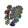
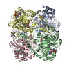
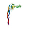
EMDB-37480: 
PSI-LHCI of the red alga Cyanidium caldarium RK-1 (NIES-2137)
PDB-8wey:
PSI-LHCI of the red alga Cyanidium caldarium RK-1 (NIES-2137)
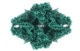
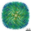
EMDB-35981:
Single-particle cryo-EM structure of mouse apoferritin at 1.49 Angstrom resolution (Dataset B)

EMDB-35984:
Single-particle cryo-EM structure of mouse apoferritin at 1.19 Angstrom resolution (Dataset A)

PDB-8j5a:
Single-particle cryo-EM structure of mouse apoferritin at 1.19 Angstrom resolution (Dataset A)

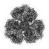
EMDB-34859:
Heteromeric ring comprised of peroxiredoxin from Thermococcus kodakaraensis (TkPrx) F42C/C46S/C205S/C211S mutant modified with 2-(bromoacetyl)naphthalene (Naph@TkPrx*F42C) and TkPrx C46S/F76C/C205S/C211S mutant modified with 2-(bromoacetyl)naphthalene (Naph@TkPrx*F76C) (Naph@(MIX|3:3))

PDB-8hla:
Heteromeric ring comprised of peroxiredoxin from Thermococcus kodakaraensis (TkPrx) F42C/C46S/C205S/C211S mutant modified with 2-(bromoacetyl)naphthalene (Naph@TkPrx*F42C) and TkPrx C46S/F76C/C205S/C211S mutant modified with 2-(bromoacetyl)naphthalene (Naph@TkPrx*F76C) (Naph@(MIX|3:3))
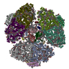
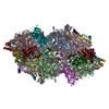
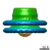
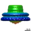
EMDB-33593:
Structure of the Anabaena PSI-monomer-IsiA supercomplex
PDB-7y3f:
Structure of the Anabaena PSI-monomer-IsiA supercomplex
EMDB-34618:
Cryo-EM structure of Beta-galactosidase at 1.57A resolution

PDB-7vi4:
Electron crystallographic structure of TIA-1 prion-like domain, A381T mutant

PDB-7vi5:
Electron crystallographic structure of TIA-1 prion-like domain, wild type sequence

EMDB-31944:
Pentacylindrical allophycocyanin core from Thermosynechococcus vulcanus

PDB-7vea:
Pentacylindrical allophycocyanin core from Thermosynechococcus vulcanus

EMDB-31945:
Phycocyanin rod structure of cyanobacterial phycobilisome

PDB-7veb:
Phycocyanin rod structure of cyanobacterial phycobilisome
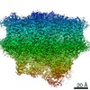
EMDB-31455:
Cryo-EM structure of a primordial cyanobacterial photosystem I
PDB-7f4v:
Cryo-EM structure of a primordial cyanobacterial photosystem I
EMDB-31062:
Structure of monomeric photosystem II

PDB-7eda:
Structure of monomeric photosystem II
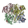
EMDB-31053:
Cryo-EM structure of Isocitrate lyase-1 from Candida albicans
PDB-7ebf:
Cryo-EM structure of Isocitrate lyase-1 from Candida albicans
EMDB-30420:
Structure of the far-red light utilizing photosystem I of Acaryochloris marina
PDB-7coy:
Structure of the far-red light utilizing photosystem I of Acaryochloris marina
EMDB-30547:
Cryo-EM Structure of PSII at 1.95 angstrom resolution
EMDB-30548:
Cryo-EM Structure of PSII at 2.08 angstrom resolution
EMDB-30549:
Cryo-EM Structure of PSII at 2.22 angstrom resolution
EMDB-30550:
Cryo-EM Structure of PSII at 2.20 angstrom resolution
PDB-7d1t:
Cryo-EM Structure of PSII at 1.95 angstrom resolution

PDB-7d1u:
Cryo-EM Structure of PSII at 2.08 angstrom resolution
PDB-7di8:
Electron crystallographic structure of Catalase using a direct electron detector at 300 kV
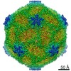
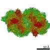
EMDB-30375:
Cryo-EM Structure of Apple Latent Spherical Virus (ALSV)

PDB-7chk:
Cryo-EM Structure of Apple Latent Spherical Virus (ALSV)
EMDB-9865:
The 1.54 A resolution structure of apoferritin by CRYOARM300 with Cold-FEG

EMDB-9890:
Cryo-EM structure of human apoferritin at 1.9A resolution by CRYO ARM 300

PDB-6jnt:
Catalase structure determined by eEFD (dataset 1)

PDB-6jnu:
Catalase structure determined by eEFD (dataset 2)

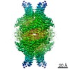
EMDB-6927:
Structure of the ExbB/ExbD hexameric complex (ExbB6ExbD3TM)

EMDB-6928:
Structure of the ExbB/ExbD pentameric complex (ExbB5ExbD1TM)

PDB-5zfu:
Structure of the ExbB/ExbD hexameric complex (ExbB6ExbD3TM)

PDB-5zfv:
Structure of the ExbB/ExbD pentameric complex (ExbB5ExbD1TM)
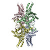
EMDB-6715:
FliF ring of Salmonella flagellar motor
EMDB-6716:
Ring assembly of FliF-FliG fusion protein
PDB-5gkn:
Catalase structure determined by electron crystallography of thin 3D crystals
PDB-3j7t:
Calcium atpase structure with two bound calcium ions determined by electron crystallography of thin 3D crystals
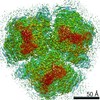
EMDB-5719:
Electron microscopy of the negatively-stained Cmr complex from Thermus thermophilus HB8.
EMDB-2418:
Structure and activity of an RNA-targeting Type III-B CRISPER-Cas complex
EMDB-1641:
Helical reconstruction of the bacterial L-type flagella filament

EMDB-1873:
The flagellar cap, HAP2 pentamer
PDB-3a5x:
L-type straight flagellar filament made of full-length flagellin
ページ:
 wwPDBはEMDBデータモデルのバージョン3へ移行します
